First magnifiers. Magnifying devices (Grade 6)
Various devices are used for biological research, many of them are technically complex and expensive. During laboratory work, it is important to properly handle instruments and tools, to follow safety rules. Always Prepare Carefully workplace to work. Place devices and equipment in such a way that they do not fall or tip over.
If we take, for example, a watermelon, an apple, an overripe cucumber or an unripe tomato fruit and break the fruit in half, we will find that inside it consists of tiny grains called cells. In order to better consider them, magnifying devices. The most common are magnifying glass and microscope.
magnifying glass
A magnifying glass is a very simple magnifying device. It consists of glass convex on both sides, usually round, and a frame where it is inserted. There are two types of magnifiers: manual and tripod. A hand magnifier can magnify the object in question by 2 to 20 times. Tripod - 10 - 25 times. Two magnifying glasses are inserted into the frame of a tripod magnifier, it also has a tripod, is equipped with an object table (for fixing the object in question) and a mirror for adjusting lighting.
Microscope
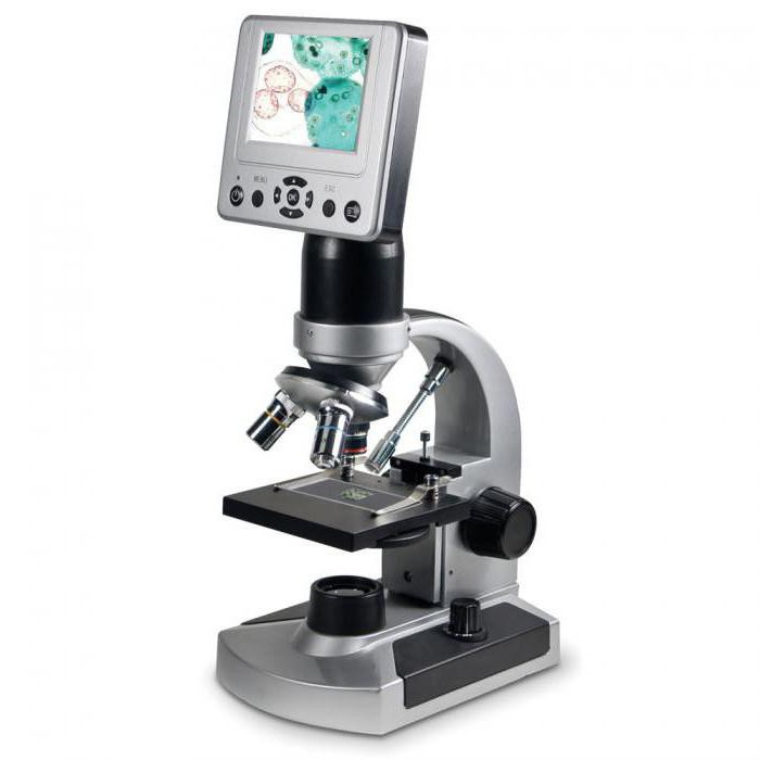
A magnifying glass allows you to see the shape of cells, but for a real study of their structure, it will not work, and here a microscope will come to the rescue. The word "microscope" itself comes from the Greek words "micros" - small and "scopeo" - look. An ordinary school microscope can magnify images up to 3600 times. Finding out how much the microscope magnifies the image is very simple, just multiply the number indicated on the eyepiece by the number indicated on the objective. If the eyepiece of a microscope gives 10x magnification, and the objective 30x, then it is enough to magnify 10 by 30 to get the number 300 - this is the total magnifying power of this device.
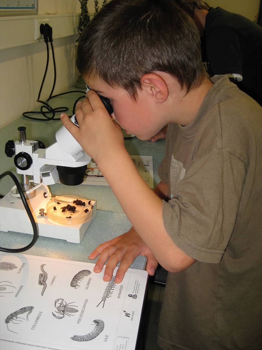
The microscope telescope contains magnifying glasses which are called lenses. At the very top of the tube, an eyepiece is screwed through which the object under study is viewed. The eyepiece usually consists of a frame and two magnifying glasses. The word "ocular" comes from the Latin "oculus", which translates as eye. The lower end of the tube is equipped with a lens, which also consists of a frame and several magnifying glasses. The word "objective" is derived from the Latin word "objectum" meaning object. The tube is attached to a tripod. An object table with a hole in the center is fixed on a tripod under it. A mirror is fixed under the object table, with which you can adjust the illumination of the object under study. Using the side screw located on the side of the tripod, adjust the distance of the lens above the object under study.
Rules for working with a microscope
- Place the microscope on the edge of the table at a distance of 5 - 15 centimeters.
- Adjust the mirror so that light enters the opening of the stage.
- Place the prepared preparation on the stage and fix it with clamps.
- Gently turning the screw, lower the tube so that it is 1 - 2 millimeters from the object.
- Look into the eyepiece, slowly turn the screw and raise the tube until a clear image of the object appears.
- Treat the microscope with care, store it in a case or a special box.
- Be sure to read the instructions carefully before using the appliance for the first time.
What other magnifying devices do modern scientists use. Prepare short message
Answer:
It is not known exactly when the first magnifiers appeared. So, during the archaeological excavations carried out on the territory Ancient Babylon, scientists found biconvex lenses - the simplest optical instruments. These lenses were made from polished rock crystal. The first microscopes invented by mankind were optical, and their first inventor is not so easy to single out and name. The earliest information about the microscope dates back to 1590 and the city of Middelburg, in Holland, and is associated with the name of Zachary Jansen, who was engaged in the manufacture of glasses. A little later, in 1624, Galileo Galilei presents his compound microscope, which he originally called "occhiolino". A year later, his friend Giovanni Faber coined the term "microscope" for the new invention. One of the first remarkable discoveries related to the improvement of magnifying instruments was made by the English scientist Robert Hooke. Robert Hooke in 1665 first saw cells (cork cut - cells - cells). A contemporary of Hooke, the Dutchman Anthony van Leeuwenhoek (1632-1723), designed a microscope that gives an increase of up to 270 times, and in the 20th century an electron microscope was invented that gives an increase of tens, hundreds of thousands of times. Huge contribution Russian scientists contributed to the development and improvement of the microscope. AT early XVIII centuries in the workshop of the Academy of Sciences in St. Petersburg, experienced specialists Kalmykov, Belyaev and others produced designs High Quality and worked fruitfully to improve these devices. High-quality microscopes were designed by the great Russian inventor IP Kulibin. Quite a lot of research with the participation of a microscope was carried out by the great Russian scientist M.V. Lomonosov. He is considered the first Russian scientist who constantly used this device in his various experiments and studies. Over time, the device of the microscope has evolved markedly, microscopes of a new type have appeared, research methods have been improved.
- What does not apply to the global problems of mankind: 1. Climate change caused by human activities 2 Gap m / y of the copper and rich strata of the population 3. Threat of pollution of the oceans 4 Threat of nuclear weapons proliferation
- Dialogue on how we spent the weekend in October conversation with a friend. Use the following words 1.Barbecue in the countryside 2.Camping in nature 3.watching videos or movies 4.going to an amusement park 5.going to the coast 6.take part in a sporting event 7.watching a sporting event 8.traveling the world 9.going to the theatre/concert/circus
Grade: Grade 6
Date: _19.09.2016
Lesson topic: " Magnifiers: microscope, magnifying glass. How to use magnifiers. Preparation of the drug. Materials and equipment. Safety. » LR №2 « Acquaintance with
Lesson type: combined
Target: formationstudents have representationsIabout the structure and principles of operation of magnifying instruments, about the significance of their invention for the development of biological sciences.
Tasks:
Educational : to form students' understanding of the structure and principles of operation of magnifying devices,aboutpractice working with magnifiers
Educational : develop logical thinking through the ability to analyze, summarize materials, draw conclusions, compare; develop observation;
Educational : bring upinterest in the subject.
Equipment: textbook "Biology" grade 6,presentation for the lesson, hand magnifier, tripod magnifier, microscope, coverslip and slide, ready micropreparation.
GreetingsPreparation for work (checking the readiness of students for the lesson)
Checking for Students
Welcome teachers.
Report dej.
Checking d / z
Oral questioning at the blackboard
Environmental factors
Plant habitat.
Answer at the blackboard
Knowledge update
The human eye is able to distinguish objects of various sizes. But there are such small structures that a person cannot see with the naked eye. How, then, have humans been able to study microscopic structures?
What magnifying devices do you know?
In what cases are they used?
Answer. Write down the date and subject.
Learning new material
The history of the microscope and the significance of this invention
First simpleThe microscope was invented in 1590 in Holland by Jansen . It is known about the device of this device that it consisted of a pipe attached to a stand with two magnifying glasses.This device was improved by another Dutchman - Anthony van Leeuwenhoek . But the first to understand and appreciate great value microscope, was an EnglishmanRobert Hooke is a physicist, meteorologist, biologist, engineer and architect. He was the first to use a microscope to study plant and animal tissues.. Robert Hooke slightly improved the microscope, and then with its help examined various objects and sketched them. One day, Hooke made a thin section of a plant cork and began to examine it under a microscope. The scientist saw that a piece of cork consists of many cells, which he called "cells". It happened in1665 As time went on, microscopes improved, scientists learned more and more about cells, their structure and functions. It turned out that not only plants, but also all other living organisms: animals, fungi, bacteria are composed of cells, i.e. have a cellular structure. modern science created super-powerful electron microscopes, allowing you to see ultra-fine structures, making it possible to study processes at the subcellular level.
Magnifier and microscope device
magnifying glass
- the simplest magnifying device. Its main part ismagnifying glass
,
convex on both sides and inserted into the frame. With the help of a magnifying glass, we see an image of an object magnified 2-25 times. The magnifying glass is taken by the handle and brought closer to the object at such a distance at which the image of the object becomes the clearest.
Microscope
- This is a device that magnifies the image of an object by several hundred and even thousands of times. The main part of the light microscope you work with at school ismagnifying glasses
,
inserted into a tube, or tube (in Latin, "tube" means "tube"). At the top end of the tube iseyepiece,
consisting of a frame and two magnifying glasses. The name "ocular" comes from the Latin word "oculus", which means "eye". When examining an object with a microscope, the eye is brought closer to the eyepiece. At the lower end of the tube is placedlens,
consisting of a frame and several magnifying glasses. The name "objective" comes from the Latin word "objectum", which means "object".
The tube is attached totripod.
Also attached to the tripodobject table
,
in the center of which there is a hole, and under itmirror.
Using a microscope, you can examine the cells of all organs of the plant. Direct the light with a mirror into the opening of the stage.
The prepared preparation is placed on the object table and the glass slide is fixed there with two clamps.
Using a screw, smoothly lower the tube so that the lower edge of the lens is at a distance of 1-2
mm from the drug.
Looking through the eyepiece, slowly raise the tube until a clear image of the object appears.
How should a microscope be used? (formulation of the rules for working with a microscope, demonstration)
Steps for preparing a preparation from a transparent onion skin (p. 21)
LR №2 " Acquaintance with magnifying instruments and laboratory instruments.
Purpose: to get acquainted with the device of magnifying devices. Learn to use a microscope.
Equipment: microscope, hand and tripod magnifier, slide and coverslip, prepared micropreparation.
Work progress:
1. Study the device of the microscope (Fig. 13)
2. Determine the sequence of preparation of the preparation from the bulb. (p. 22 "I")
3. Define functions constituent parts microscope (p. 23 table)
Page 22 "I"
Cover the preparation with a cover glass - 5
Peel the onion from the husk - 2
Remove the thin skin from the convex side of the bulb - 3
Put some water on the glass slide - 1
A thin skin is straightened with a dissecting needle - 4
Install the drug on the subject table - 6
Page 23 table
Listen to the teacher's story
Studying the structure of a microscope
Perform laboratory work
Anchoring
1. What magnifying devices do you know?
2. What is a loupe and how much magnification does it give?
3. How does a microscope work?
4. How do you know what magnification a microscope gives?
Answer questions
Reflection
Outcome
Reflection
I found out…
I like it…
I was having a hard time...
Grading
Summing up the lesson.
D/ h
draw up an LR
Write down d.z.
Afanasyeva N.V.
5th grade high school is the time when we first get to know them. In the lessons, children are told the most basic things about their device and creators. Would you like to deepen your knowledge about them? Or maybe you are preparing a lesson on the topic "Magnifying Instruments" (Grade 5)? In any case, we have something to tell you.
ancient lenses
The history of the discovery of magnifying devices begins in the distant past. A large plano-convex lens has come down to us - one of the most ancient. Its diameter is 55 mm, and focal length- about 150 mm. It was made of rock crystal for 2.5 thousand years BC. e. It was discovered in 1890 by G. Schliemann during the excavations of Troy. Around 600-400 years. BC e. started making glass lenses. They were discovered in Sargon (this is Mesopotamia). In Sweden in 1877 a double lens 5 cm in diameter was found, convex on both sides. It dates back to 500 AD. e. You can continue the list of ancient lenses that researchers have discovered for a long time. The history of the discovery of magnifying devices has many facts. Despite this, one can only speculate about how they were used at that time.
Contribution of Roger Bacon
Modern scientists have familiarized themselves with a thorough description of lenses made by Roger Bacon (years of life - 1214-1294). He was a graduate of Oxford University and also became famous as a prominent thinker and scientist. Lenses, according to his work, were used to magnify the image. From the translation of a fragment of the work, it follows that Bacon was able to correctly describe the effect of lenses that served as a reverse telephoto lens (we are talking about the description of a single-component optical tube).
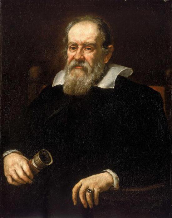
Merit of Galileo Galilei
The history of the discovery of magnifying devices is unthinkable without the name of this man. About 300 years after Bacon's death, Galileo Galilei, a famous scientist from Italy, created a similar pipe. It was not three-, but two-component. Virtually a "peer" of this is a microscope. It is generally accepted that he owes his appearance to Galileo. Galileo parted the telescope and noticed that small objects in this state can be well enlarged. D. Viviani confirms that it was Galileo who invented the microscope. Viviani, by the way, wrote a biography of this Italian scientist.
An important event for science was the discovery of magnifying instruments in 1625. It was then that Faber, a member of the Roman Academy, first used the term "microscope" in relation to the invention made by Galileo.
What was created by Drebel and Alkmar, the development of Tore and Hooke
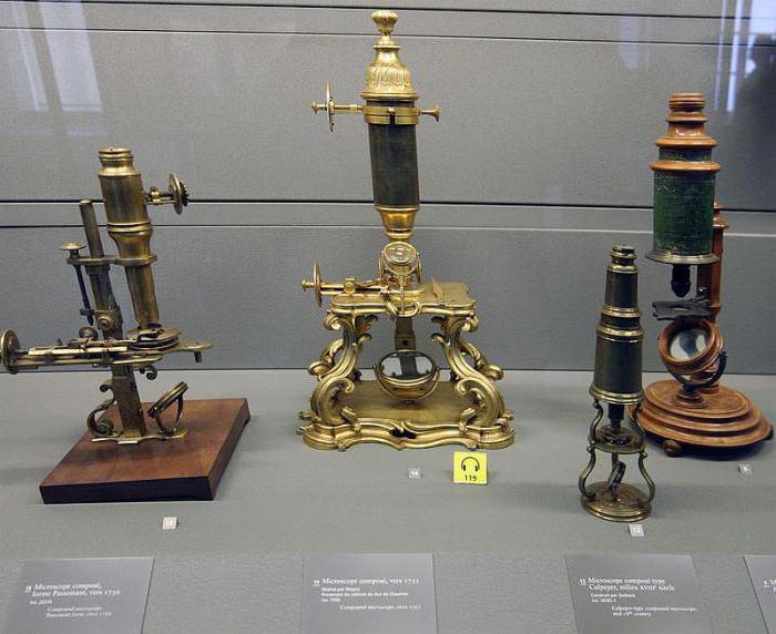
The history of the discovery of the microscope continues with the work of K. Drebel and Alkmar. These Dutch scientists designed a device that consisted of two convex lenses. Due to this, the image of the object that was viewed under it was presented upside down. This compound microscope, which had a biconvex or plano-convex eyepiece, as well as a biconvex objective, is considered the forerunner of later compound microscopes (one of them is shown in the photo below).

The Italian Tore around 1660 made spherical magnifiers from frozen drops of glass. The history of the discovery of the microscope is inconceivable without this name, since the magnifiers created by the Italian made it possible to magnify objects one and a half thousand times.
Does another name tell you anything - Robert Hooke? This English scientist made a great contribution to the discovery of magnifying instruments. Robert Hooke improved them so much that it became one of significant events in the history of optics. Hooke's microscope diagram is shown in the photo below.

Thanks to this invention, in 1665, Robert was able to see cells for the first time on a section of cork. Thus, such a science as biology received an important technical tool. Magnifying devices continued to improve Leeuwenhoek. Let's talk about him.
Leeuwenhoek and his achievements
A significant contribution to the history of the development of magnifying instruments was made by A. V. Leeuwenhoek, a Dutchman who lived in a city like Delft. The years of his life - 1632-1723. He independently designed and used simple microscopes in research (one of the models of such devices is presented below), capable of magnifying up to three hundred times.
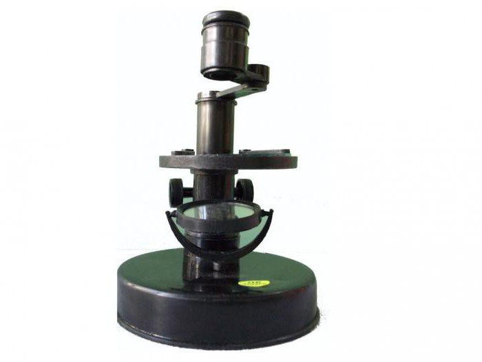
It was Leeuwenhoek who first compiled a description of microscopic organisms (including unicellular bacteria), based on his observations. In 1698, Peter I, the Russian Tsar, paid a visit to this famous explorer. Peter was in Holland at that time and, as you know, was interested in everything new. For his Kunstkamera, opened by him in St. Petersburg, he purchased several complex and simple microscopes. And much later, after the opening of the Academy of Sciences, they were placed at the disposal of this organization.
Works of Russian scientists from the Academy of Sciences
The lesson "Magnifying Instruments" should also include a story about the achievements in optics of representatives of our country. Promising Russian scientists, whose work was led by M. V. Lomonosov, began to use microscopes bought by Peter I in biological research. And later they actively participated in their improvement.
The discovery of magnifying instruments continued in 1747. It was then that L. Euler, a member of the Academy of Sciences of St. Petersburg (years of life - 1707-1783), suggested using an achromatic lens for a microscope. The fundamental work of this scientist in the field of geometric optics is "Dioptric". It consists of three volumes, which were published in 1769-1771. A new microscope, already achromatic, was released in 1802, after the work of Elinus (also a member of the Academy of Sciences of St. Petersburg) was published.
Such a microscope at that time was considered perfect to such an extent that scientists did not even allow the thought that it could be improved. This discovery made a lot of noise at the time. The arrangement of Elinus' magnifying instruments was as follows. They were equipped with six lenses, it was possible to change the magnification smoothly, the distance from the object to the image changed. It was in our country that the idea of an achromatic microscope with variable magnification, important for science, was born and brought to life. However, this idea did not take root in further developments. Changing the magnification of the instrument by adjusting the length of the tube, however, was an important idea that made a significant contribution to the history of development Today, one of the microscopes created by Elinus can be seen in Polytechnic Museum Moscow, which belongs to the Institute of History, Natural Science and Technology. The photo below shows magnifying instruments dating back to the 18th century.
Further improvement of microscopes
J. G. Tiedemann, a German optician from Stuttgart, set about building two achromatic microscopes in the early 19th century. The University of Derpt (today it is called Tartu) gave him cash for the implementation of work. In 1808, these devices were released.
In 1807, a year before the development of achromatic microscopes, Van Dale, a Dutch optician, published his work. It presented a description of the design of an achromatic microscope created by him. Western European historians believe that the microscope created by this particular scientist was the first such device of satisfactory quality. However, in all respects it was inferior to the one designed by Elinus. By the way, the achromatic microscopes of I. Fraunhofer, released in 1811, were distinguished by an even more imperfect design when compared with the microscopes of Elinus.
Russian microscopes in the 19th century
In the first half of the 19th century, magnifying instruments were already produced in many places on earth. In Russia, their production began in the 18th century, but subsided by the beginning of the 19th century. It is known that around 1820 microscopes of rather high quality were produced by an optics workshop located at Kazan University. However, in Russia there was still no rapid development of this industry, since the government of that time believed that the best option was to buy magnifiers abroad.
Contribution to optics by Giambattista and Amici
Amici Giambattista (years of life - 1786-1863) - a famous Italian optician, astronomer and botanist. He devoted many years of his life to the development of microscopy. In 1827, Amici himself designed and made an achromatic lens with an aperture of 0.60 and good aberration correction. The same scientist in 1844 began experiments on the use of water and oil immersion. Thanks to them, the production of objectives with a numerical aperture of 1.30 and water immersion was launched.
Abbe microscopes
Oil immersion instruments with an aperture of 1.50 (which are still in use today) began to be produced thanks to the work of Ernst Abbe, a German optician. He invented the law of sines, with the help of which the coma observed in small linear fields was eliminated. E. Abbe continued to develop the theory of image formation in a magnifying device. He also clarified the issue of these devices. Abbe was the head of the work on the creation of a whole series of high quality achromatic microobjectives. Their numerical aperture reached 1.50. These devices were produced in Jena by the company "K. Zeiss" (in 1872). The same company under the leadership of E. Abbe made 8 apochromats. And in 1888, its employees developed an apochromat, which had an aperture of 1.60 and had monobromonaphthalene immersion.
Recent major advances in optics
Russian scientists D. S. Rozhdestvensky and L. I. Mandelstam developed Ernst's theory in their works. An important merit of Rozhdestvensky was that he introduced the concept of relative incoherence of illumination. R. Richter, an employee of the K. Zeiss company, developed and received a patent for a special lighting device used in a microscope. However, the problem of the optimal ratio of the parameters of interchangeable lenses and the lighting system is still relevant today. Domestic microscopes today are in no way inferior in terms of technical performance and optical parameters to devices created by famous companies Abroad.
So, we briefly outlined the history of the emergence of modern microscopes. When developing the lesson "Magnifying Instruments" (Grade 5), you can use the information presented in the article.


, poultry farming")


- Burns, Robert - short biography
- The concept of common vocabulary and vocabulary of limited use
- Nancy Drew: The Captive Curse Walkthrough Nancy Drew Curse of Blackmoore Manor Walkthrough
- Deadpool - Troubleshooting
- Won't start How to Survive?
- What to do if bioshock infinite won't start
- Walkthrough Nancy Drew: Alibi in Ashes
- Spec Ops: The Line - game review, review Spec ops the line crashes on missions
- Room escape level 1 walkthrough
- Processing tomatoes with boric acid How much will 2 grams of boric acid
- Cucumber Grass (Borago)
- Bioinsecticide Lepidocid: purpose, properties and application procedure Lepidocide waiting period
- How to change the language to Russian in steam
- Dendrobium noble: room care
- Morphology of plants general concepts - document
- Planting, propagation and care of bamboo at home, photo Growing bamboo from seeds
- How to strengthen the cellular signal for the Internet in the country
- Sanskrit reveals the forgotten meaning of Russian words (2 photos)
- The oldest language Sanskrit programming language of the future Dead language Sanskrit
- Who has dominion over all the earth?