Microscope. The principle of operation of the microscope is the construction of the image. Microscope and microscopic research methods
Microscope(from Greek. mikros- small and skopeo- look) - optical instrument to obtain an enlarged image of small objects and their details, invisible to the naked eye.
The first known microscope was created in 1590 in the Netherlands by hereditary opticians Zachary and Hans Jansenami who mounted two convex lenses inside one tube. Later Descartes in his book "Dioptrics" (1637) he described a more complex microscope, composed of two lenses - a plano-concave (eyepiece) and a biconvex (objective). Further improvement of optics allowed Anthony van Leeuwenhoek in 1674 to make lenses with a magnification sufficient to carry out simple scientific observations and for the first time in 1683 to describe microorganisms.
A modern microscope (Figure 1) consists of three main parts: optical, illumination and mechanical.
Main details optical part microscope are two systems of magnifying lenses: the eyepiece facing the eye of the researcher and the lens facing the preparation. Eyepieces They have two lenses, the upper of which is called the main, and the lower collective. On the frame of the eyepieces indicate what they produce increase(×5,×7,×10,×15). The number of eyepieces in the microscope may be different, and therefore distinguish monocular and binocular microscopes (designed to observe an object with one or two eyes), as well as trinoculars , allowing you to connect to the microscope documentation systems (photo and video cameras).
Lenses They are a system of lenses enclosed in a metal frame, from which the front (frontal) lens produces an increase, and the corrective lenses lying behind it eliminate the imperfections of the optical image. On the frame of the lenses, the numbers also indicate what they produce. increase (×8,×10,×40,×100). Most models designed for microbiological research are equipped with several lenses with different magnifications and a rotary mechanism designed for quick change - turret , often called " turret ».
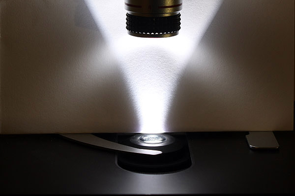
lighting part is designed to create a light flux that allows you to illuminate the object in such a way that the optical part of the microscope performs its functions with the utmost accuracy. The illuminating part in a direct transmitted light microscope is located behind the object under the lens and includes Light source (lamp and electrical power supply) and opto- mechanical system (condenser, field and aperture adjustable diaphragms). Condenser consists of a system of lenses that are designed to collect rays coming from a light source at one point - focus , which must be in the plane of the object under consideration. In its turn d diaphragm located under the condenser and designed to regulate (increase or decrease) the flow of rays passing from the light source.
Mechanical The microscope contains parts that combine the optical and illumination parts described above, as well as allowing you to place and move the specimen under study. Accordingly, the mechanical part consists of grounds microscope and holder , to the top of which are attached tube - a hollow tube designed to accommodate the lens, as well as the turret mentioned above. Below is object table on which glass slides with test specimens are placed. The stage can be moved in the horizontal plane using the appropriate device, as well as up and down, which allows you to adjust the sharpness of the image using coarse (macrometric) and precision (micrometric) screws.
Increase, which gives the microscope is determined by the product of the magnification of the objective and the magnification of the eyepiece. In addition to light-field microscopy, dark-field, phase-contrast, luminescent (fluorescent) and electron microscopy have been widely used in special research methods.
Primary(own) fluorescence occurs without special treatment of drugs and is inherent in a number of biologically active substances, such as aromatic amino acids, porphyrins, chlorophyll, vitamins A, B2, B1, some antibiotics (tetracycline) and chemotherapeutic substances (akrihin, rivanol). Secondary (induced) fluorescence arises as a result of processing microscopic objects with fluorescent dyes - fluorochromes. Some of these dyes are diffusely distributed in cells, while others bind selectively to certain cell structures or even to certain chemicals.
For this type of microscopy, special fluorescent (fluorescent) microscopes , which differ from a conventional light microscope in the presence of a powerful light source (Ultra-high pressure mercury-quartz lamp or halogen quartz incandescent lamp), which emits predominantly in the long-wave ultraviolet or short-wave (blue-violet) region of the visible spectrum.
This source is used to excite fluorescence before the emitted light passes through a special exciting (blue-violet) light filter and reflected interference beam-splitting plate , which almost completely cut off longer wavelength radiation and transmit only that part of the spectrum that excites fluorescence. At the same time, in modern models In fluorescent microscopes, the excitation radiation enters the preparation through the objective (!) locking (yellow) light filter , which cuts off short-wave exciting radiation and transmits luminescence light from the preparation to the observer's eye.
Due to the use of such a system of light filters, the luminescence intensity of the observed object is usually low, and therefore luminescence microscopy should be carried out in special darkened rooms .
An important requirement when performing this type of microscopy is also the use of non-fluorescent immersion and confining media . In particular, to quench the intrinsic fluorescence of cedar or other immersion oil, small amounts of nitrobenzene are added to it (from 2 to 10 drops per 1 g). In turn, a buffer solution of glycerol, as well as non-fluorescent polymers (polystyrene, polyvinyl alcohol) can be used as concluding media for preparations. For the rest, when conducting luminescence microscopy, conventional slides and coverslips are used, which transmit radiation in the part of the spectrum used and do not have their own luminescence.
Accordingly, the important advantages of fluorescent microscopy are:
1) color image;
2) a high degree of contrast of self-luminous objects against a black background;
3) the possibility of studying cellular structures that selectively absorb various fluorochromes, which are specific cytochemical indicators;
4) the possibility of determining functional and morphological changes in cells in the dynamics of their development;
5) the possibility of specific staining of microorganisms (using immunofluorescence).
electron microscopy
The theoretical foundations for using electrons to observe microscopic objects were laid W. Hamilton , who established an analogy between the passage of light rays in optically inhomogeneous media and particle trajectories in force fields, and also de Broglie , who put forward the hypothesis that the electron has both corpuscular and wave properties.
At the same time, due to the extremely short electron wavelength, which decreases in direct proportion to the applied accelerating voltage, the theoretically calculated resolution limit , which characterizes the ability of the device to display separately small, as close as possible details of the object, for an electron microscope is 2-3 Å ( angstrom , where 1Å=10 -10 m), which is several thousand times higher than that of an optical microscope. The first image of an object formed by electron beams was obtained in 1931. German scientists M. Knolem and E. Ruska .
In the designs of modern electron microscopes, the source of electrons is a metal (usually tungsten), from which, after heating to 2500 ºС, as a result thermionic emission electrons are emitted. With the help of electric and magnetic fields, the emerging electron flow you can speed up and slow down, as well as deflect in any direction and focus. Thus, the role of lenses in electron microscope plays a set of appropriately calculated magnetic, electrostatic and combined devices, called " electronic lenses" .
A necessary condition for the movement of electrons in the form of a beam on long distance is also a creation on their way vacuum , since in this case the mean free path of electrons between collisions with gas molecules will significantly exceed the distance over which they must move. For these purposes, it is sufficient to maintain a negative pressure of approximately 10 -4 Pa in the working chamber.
By the nature of the study of objects, electron microscopes are divided into translucent, reflective, emissive, raster, shadow and mirror , among which the first two are the most commonly used.
Optical design transmission (transmission) electron microscope is completely equivalent to the corresponding optical microscope design, in which the light beam is replaced by an electron beam, and glass lens systems are replaced by electronic lens systems. Accordingly, a transmission electron microscope consists of the following main components: lighting system, object camera, focusing system and final image registration unit consisting of a camera and a fluorescent screen.
All these nodes are connected to each other, forming the so-called “microscope column”, inside which a vacuum is maintained. Another important requirement for the object under study is its thickness less than 0.1 µm. The final image of the object is formed after the appropriate focusing of the electron beam passed through it on photographic film or fluorescent screen , coated with a special substance - a phosphor (similar to the screen in TV kinescopes) and turning the electronic image into a visible one.
In this case, the formation of an image in a transmission electron microscope is mainly associated with a different degree of electron scattering by different parts of the sample under study and, to a lesser extent, with a difference in the absorption of electrons by these parts. The contrast is also enhanced by applying " electronic dyes "(osmium tetroxide, uranium, etc.), selectively binding to some parts of the object. Arranged In a similar way Modern transmission electron microscopes provide maximum useful magnification up to 400,000 times, which corresponds to resolution at 5.0 Å. The fine structure of bacterial cells revealed using transmission electron microscopy is called ultrastructure .
AT reflective (scanning) electron microscope The image is created by electrons reflected (scattered) by the surface layer of an object when it is irradiated at a small angle (approximately a few degrees) to the surface. Accordingly, the formation of an image is due to the difference in the scattering of electrons at different points of the object, depending on its surface microrelief, and the result of such microscopy itself appears as a structure of the surface of the observed object. Contrast can be enhanced by spraying metal particles onto the object's surface. The achieved resolution of microscopes of this type is about 100 Å.
optical microscope - a device for obtaining enlarged images of objects (or details of their structure), invisible naked eye. (from other Greek. μικρός "small" and σκοπέω "I examine") - an optical device for obtaining enlarged images of objects (or details of their structure) invisible to the naked eye. Source: Wikipedia.
Fields of application of microscopes
Optical microscopes vary in types and modifications for a variety of applications.
Microscopy methods in modern world used in almost all areas human activity: "list areas of use"
In recent decades, special optical software has been widely used for microscopic studies. By using computer programs continuous observation of research objects is achieved, which is especially important for the study of biological objects.
Thanks to modern algorithms used in optical software, labor costs are significantly reduced
Device principles
The main components of a microscope are:
The optical microscope system includes a number of components, the main of which is the lens.
The optics of the microscope consists of two elements - an eyepiece and an objective, which are fixed in a movable tube, located on a metal base with an object stage. The magnification of a microscope without additional lenses between the eyepiece and the objective is equal to the product of their magnifications
Nowadays, a microscope almost always has an illumination system and micro and macro screws to adjust the sharpness.
Depending on the purpose, additional systems and devices may be attached to the research microscope, such as
objectives with increased resolution 40, aperture 0.65, correction for the thickness of the coverslip 0.17 mm and infinite length of the tube
Optical microscope objectives are one of the main parts and are a complex mechanism for enlarging the image of the subject under study. An image of an object magnified with an optical lens is viewed through an eyepiece, which in turn can also create magnification. If the microscope lens somehow distorts the image, then this distortion will be amplified by the eyepiece. A microscope objective is a complex optical system that magnifies the image of an object. It is the most responsible and main part of the research equipment. You can view the image created by the lens through the eyepiece.
The objectives of research and other microscopes, including stereoscopic ones, are mostly interchangeable and unified. The interchangeability is primarily affected by the lens mounting parameters.
The objects of microscope research can be any organic and non-organic objects, living and non-living tissues, whole biological organisms or their separate parts.
The microscope has as an illuminating optical system halogen lamp or LED system. The advantage of the LED is an extremely long operating time compared to conventional halogen lamps (100 or more times longer than this indicator); low power consumption (constituting from 1/3 to 1/10 of the energy consumption of a conventional lamp); spectral “purity”, etc.
Capacitors

Optical microscope condensers are the main element of the system and for the most part are a separate, more often - removable, unit. The condensers are mounted directly next to the object stage and illuminate the object. An integral part of the condenser is the aperture iris diaphragm.
The diaphragm is designed to limit the amount of light to only that part of the preparation that is being studied in this moment time. This is especially useful when working at high magnifications, when only a small area of the specimen needs to be illuminated.
An open field diaphragm increases the width of the light beam. This setting is used when working at low magnifications (larger field of view)

Closing the aperture narrows the beam of light
Microscope stage
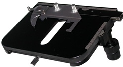
An integral part of the design that the microscope has is object table, which is the surface on which the drug is placed for research. Subject tables are divided into movable and fixed. Fixed object tables are mounted on the simplest and cheapest equipment used to teach children in schools.
Even the simplest microscope stages allow movement in two coordinate planes, and more complex ones provide movement along three axes and rotation at a certain angle.
Applicable lenses and their main characteristics
As mentioned earlier, optical microscopes whose lenses are one of the most important parts. This is a highly complex optical design that integrates a front lens and a combination of internal lenses. Depending on the level of tasks assigned, the lens can have up to fourteen different lenses.
The main data is usually indicated on the body of the optical lens.
The microscope may have the following objectives:
- Achromats (achromatic);
- Planpochromatic
- Planachromatic
- Planfluorates
Achromatic the lenses correct the aberration of the red and violet spectra. They also reduce spherical aberration, spherochromatic aberration.
Planachromatic lenses almost completely eliminate spherical aberration. Unlike achromatic lenses, apochromatic ones almost do not distort the natural color of the object.
Main advantage planpochromatic optical lenses is the ability to use them to get a sharp and not distorted image across the entire field. In addition, some modifications of flat field lenses correct chromatic aberrations.
The degree of magnification of the image of the object under study is one of the main parameters of optical lenses. According to the degree of magnification, lenses are divided into:
- low magnification - up to 10x;
- medium magnification - from 10x to 50x;
- high magnification - from 50x to 100x;
Next important characteristic objectives is their numerical aperture, which shows the resolution of the optical system of the microscope and is determined by the value of the minimum distance at which the lens can distinguish between two adjacent points.
Objectives are classified according to aperture size.
- lenses with a small aperture - up to 0.25;
- with an average aperture - up to 0.65;
- with a large aperture - more than 0.65.
Nikon Microscopes
branded microscopes Nikon occupy the highest rank. These are modern microscopes, in which the designers have integrated the latest and most innovative technical solutions and the possibilities of world science and technology.
By field of application company microscopes Nikon are divided into the following groups:
- biological microscope;
- stereomicroscopes.
Biomedical or biological microscopes Nikon are used for modern biological and medical research on living organisms and objects, as well as for automated and multi-purpose laboratory analyses.
Among biomedical Nikon the following model lines are distinguished:
- Microscope Nikon Eclipse E;
- Microscope Nikon Eclipse Ci;
- Nikon microscope Ni;
- Nikon microscope Ti.
Stereomicroscopes Nikon allow the operator to observe a three-dimensional object of study with the possibility of obtaining a completely natural image.
Among the Nikon stereomicroscopes, the following series of models stand out:
- Microscope Nikon SMZ1270/1270i;
- Microscope Nikon SMZ800N;
- Microscope Nikon SMZ25/SMZ18;
- Microscope Nikon SMZ745/745T;
- Nikon SMZ 660;
- Nikon SMZ 445/460.
Image documentation.
Integration of modern microscopes Nikon with digital cameras allows you to continuously monitor the objects under consideration with simultaneous capture and recording of their images. Digital cameras are currently widely used for observing living organisms, as well as in other branches of science and technology.
Nikon manufactures the following digital cameras:

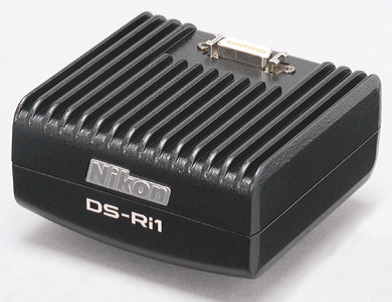
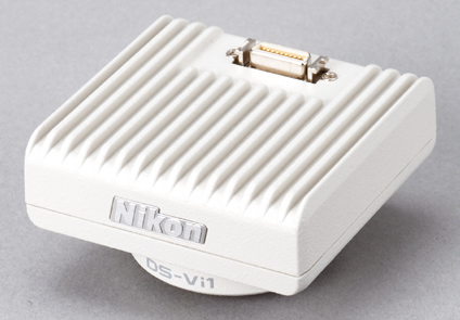

Nikon DS-Fi2 Nikon DS-Qi1 Nikon DS-Vi1 Nikon DS-Fi1c Nikon DS-Ri1
- digital camera NikonDS-Fi2;
- digital camera NikonDS-Qi1;
- digital camera NikonDS-Vi1;
- digital camera NikonDS-Fi1c;
- digital camera NikonD.S.- Ri1 .
Microscope catalog
| Straight microscopes | Eclipse E |
| Eclipse Ci | |
| NikonNi | |
| NikonTi | |
| Stereomicroscopes | SMZ25/SMZ18 |
| SMZ745/745T | |
| SMZ800N | |
| SMZ660 | |
| SMZ445/460 |
Classification according to the principle of imaging
In laboratory microscopes, the observer does not always see reflected or transmitted light as if he were looking with the naked eye. The beam of light may be subject to change, both in shape and in wavelength or other properties. In this regard, there are several types of laboratory microscopes according to the principle of imaging:
- bright field method. For ordinary person this is the most convenient form of object perception: a light background and a dark image. Used in transmitted light microscopes, so the observer gets the same image but magnified. Changes can only be caused by the use of colored glass filters that are put on the lens. Less commonly used are interference filters that only allow a certain wavelength to pass through.
- Dark field method. In these microscopes, the opposite is true: a dark background and more light image or a bright shiny contour of the object under study. This is achieved in different ways, depending on the type of microscope. In transmitted light, the incident light is blocked until it hits the object. In devices of reflected light, the beam passes through an annular diaphragm with an opaque disk, which is larger than the exit pupil of the lens.
- Phase contrast method. These microscopes, sometimes referred to as phase microscopes, make it possible to obtain images with clearly defined external and internal boundaries. This method is well suited for studying cells and tissues.
- Luminescent microscopes. Their principle of operation is based on the properties of certain substances to excite their own radiation under the action of ultraviolet or blue-violet rays. An appropriate bright light source is directed at the object, and new rays from it are "cut off" by a complex system of light filters until only a certain wavelength of radiation is obtained.
- Immersion microscopes. These devices are used for complex biomedical research, where it is necessary to obtain a contrast image of an object against a background of a similar shade. Direct transmitted light is blocked in two stages: part before the object, the second part - after the object with attenuation.
- Microscopes of interference (or differential interference) contrast. Allow to receive plain background volumetric image of the same color. A border of a different color is used to separate the image from the background.
- Ultraviolet and infrared microscopes. In them, illumination and image formation occurs at wavelengths invisible to the human eye. Accordingly, for the convenience of observations, such microscopes are connected to a computer that converts the image.
Modern laboratory microscopes are not always built according to any one principle. It is economically unprofitable for a laboratory to purchase dozens of instrument models for various observations, so now microscopes are produced in a modular design to form different ways imaging. In addition, many can be connected to a computer for recording and processing information.
Classification by lighting method
For getting quality results Observations must be made in good light. Natural light is used only by toy or school microscopes, and for laboratory instruments additional light sources are needed. Depending on their type and location in the microscope system, the following design options are distinguished:
- Transmitted light microscopes. The standard way of building a microscope, which was used in the very first models and is often found today. The principle of their operation is related to the fact that light from an external source passes through the object, and at this time a person observes it through a binocular nozzle. Microscopes of all types, including stereoscopic ones, can be built according to this principle. With their help, you can study transparent and translucent objects.
- reflected light microscopes. Here the observer does not see the object of study directly, but looks at the image that is reflected from it. Flat field microscopes (inverted or straight) as well as stereoscopic microscopes can be manufactured according to this principle. With the help of reflected light, it is good to examine opaque objects with varying degrees reflectivity, as well as translucent samples.
In turn, laboratory reflected light microscopes are also divided into two main categories:
- The "original" reflected light microscopes, in which light passes through the optical system of the microscope, reflects off the object, and then passes through the optics again. In the first case, the lens becomes part of the lighting system, in the second - the main element that increases the light reflected from the object and transmits it to the observer.
- In the second version of the design, the light falls on the object directly, and not through the optical system of the microscope. Magnification occurs due to the passage of reflected light through the lens. Stereoscopic microscopes are usually built according to this principle.
There are also flat-field luminescent devices in which there is a reflected light illuminator. In them, the image under consideration is not built by the beam of light that passed through the optics, reflected from the object and again passed through the lens. In other words, the same beam of light is used, but its length after reflection from the object and re-passing through the optics will be different. It often happens that different lighting systems are combined in one microscope. This is done in order to make the device universal for studying all kinds of objects.
Target: get acquainted with the structure of the microscope, the rules for working with it, the technique for making simple preparations, the rules for processing the results of observations.
Materials and equipment: microscope, slides and coverslips, droppers with water and lactophenol, dissecting needles, club moss spores, mallow pollen, begonia leaf petioles, tradescantia leaves.
The structure of the microscope
A microscope is an optical-mechanical device that allows you to get a greatly enlarged image of the object in question, the dimensions of which lie beyond the resolution of the naked eye. A person with normal vision distinguishes two points as two or two lines as two, and not one, only if the distance between them is at least 100 microns. Thus, the resolving power of the eye is low. When working with a microscope, the distance between two points or lines, at which they do not seem to merge, is reduced to tenths of a micrometer. In other words, the resolution of light microscopes is 300-400 times higher than the resolution of the naked eye and is equal to 0.2-0.3 microns.
The useful magnification of modern optical microscopes reaches 1400 times, while revealing the smallest details of the structure of the object under study.
In a microscope, optical and mechanical systems are distinguished.
The optical system consists of three parts: an illuminator, an objective, and an eyepiece (Fig. 1).
A tube is located between the objective and the eyepiece. All these parts are strictly centered and mounted in a tripod, which is the mechanical system of the microscope. The tripod consists of a massive base, an object table, an arc, or a tube holder, and feed mechanisms that move the object table in a vertical direction.
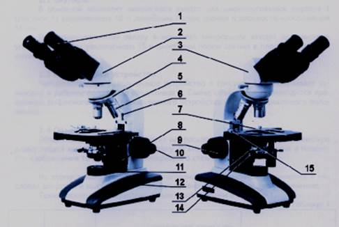
Rice. 1. Light monocular device (A)
and binocular (B) microscope:
1 - eyepieces; 2 - binocular attachment; 3 – nozzle fastening screw; 4 - revolving device; 5 - lenses; 6 - screw stop (limiter of movement of the object table during focusing; 7 - object table; 8 - handle for moving the object table in two mutually perpendicular directions; 9 - coarse focusing handle; 10 - fine focusing handle; 11 - collector in the frame; 12 - base of the microscope; 13 - condenser; 14 - condenser fixing screw; 15 - preparation parent
The lighting apparatus is represented by a condenser with an iris diaphragm and an illuminator with a halogen incandescent lamp. The condenser is located in a ring under the microscope stage. It consists of two or three lenses inserted into a cylindrical frame. The condenser serves for the best illumination of the study drug. The front lens of the condenser should be installed at the level of the microscope stage or slightly below it.
At the bottom of the condenser is an iris diaphragm. It is a system of numerous thin plates ("petals"), movably fixed in a round frame. Using the adjusting ring, you can change the size of the diaphragm opening, which always maintains a central position. This regulates the diameter of the beam of light coming from the lamp into the condenser. A ring is fixed under the diaphragm, into which a light filter is inserted, usually made of frosted glass.
The illuminator built into the base of the microscope includes a collector in a frame, which is screwed into the hole in the base, and a holder for a 6V, 20W halogen incandescent lamp. The illuminator is turned on using a switch located on the rear surface of the microscope base. By rotating the lamp incandescence adjustment dial, located on the side surface of the microscope base to the left of the observer, one can change the brightness of the lamp incandescence.
Having passed through the condenser and refracted in its lenses, the rays coming from the light source illuminate the specimen lying on the microscope stage, pass through it, and then enter the lens in the form of a divergent beam.
By partially covering the lower lens of the condenser, the diaphragm blocks side rays, resulting in more sharp image object.
The lens is the most important part of the optical system. It consists of several lenses set into a metal sleeve. High magnification lenses include 8–10 lenses or more. The lens gives an image of the object with the reverse arrangement of parts. In doing so, it reveals ("resolves") structures that are inaccessible to the naked eye, with greater or lesser detail, depending on the quality of the lens. The image is built by the lens in the plane of the aperture of the eyepiece located in the upper part of the tube (tube) of the microscope. The optical properties of a lens depend on its design and the quality of the lenses. The most powerful lenses give 120x magnifications. On the laboratory classes usually work with lenses that magnify 4, 20, 40 times.
Great importance when working with a microscope, it has the working distance of the objective, i.e. the distance from the lower (front) lens of the objective to the object (to the upper surface of the slide). For lenses with 40x magnification, this distance is 0.6 mm. Therefore, it is desirable to use coverslips that are thinner than the working distance. The normal thickness of the coverslip is 0.17–0.18 mm.
The eyepiece is much simpler than the lens. Some eyepieces consist of only two lenses and a diaphragm inserted into a cylindrical frame. The upper (ocular) lens serves for observation, the lower ("collective") plays an auxiliary role, focusing the image built by the lens. The aperture of the eyepiece defines the boundaries of the field of view.
At the lower end of the tube holder, a revolving device is fixed - a rotating disk with sockets that have threads for screwing in lenses. The screw threading of the turret sockets and objectives is standardized, so the objectives fit microscopes different models. The tube holder is fixedly connected to the tripod.
The microscope is designed so that the preparation is located between the main focus of the objective and its double focal length. In the microscope tube, in the plane of the eyepiece diaphragm, located between the main focus and the optical center of the upper lens of the eyepiece, the objective builds a real magnified inverse image of the object. Acting like a magnifying glass, the top lens or eyepiece lens system produces a virtual upright magnified image. Thus, the image that is obtained using a microscope turns out to be twice enlarged and inverse with respect to the object under study (Fig. 2). The total magnification of a microscope with a normal (160 mm) tube length is equal to the magnification of the objective multiplied by the magnification of the eyepiece.
The square stage has a hole in the center, into which the top of the condenser fits. The object table together with the preparation can be moved back and forth. Modern microscopes are also equipped with a preparation guide, with which the preparation can be moved back and forth on the stage. For this, two screws are located on the axis on the right.
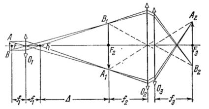
Rice. 2. The path of the rays in the microscope:
AB - subject; O 1 is a microscope lens that gives an enlarged inverse and real image of the object A 1 B 1 . The image of the object lies in the focal plane F 2 of the microscope eyepiece O 2 , through which it is viewed as through a magnifying glass. In the focal plane F 3 of the lens of the eye O 3 a real image of the object A 2 B 2 is obtained. Such an arrangement of O 1 and O 2 is also possible, when A 1 B 1 is located between F 2 and O 2
under the subject table. The upper screw is used to move the object table, and the lower screw is used to move the preparation.
The movement of the drug with the object for sharpening is carried out by moving the object table, which is movably connected to the tube holder. With the help of feed mechanisms, it can be moved vertically (up - down) to focus. In most modern microscopes, these mechanisms (screws) are fixed at the base of the tube holder.
Coarse focusing is carried out using a macrometric screw (kremalery). Fine focusing is carried out with a micrometer screw. Divisions are applied on the drum of the micrometer screw. A movement of one division corresponds to raising or lowering the pipe by 2 µm. With a full turn of the screw, the pipe moves 100 µm.
Mechanisms for macrometric and especially micrometric feeds are made very precisely and require careful handling. Rotate the screws should be smooth, without jerks and force.
There are various models of educational and research light microscopes. Such microscopes make it possible to determine the shape of microorganism cells, their size, mobility, the degree of morphological heterogeneity, as well as the ability of microorganisms to differentiate staining.
The success of observing an object and the reliability of the results obtained depend on a good knowledge of the optical system of the microscope.
Consider the device and appearance of a biological microscope, model XSP-136 (Ningbo teaching instrument Co., LTD), its operation constituent parts. The microscope has mechanical and optical parts (Figure 3.1).
Figure 3.1 - Device and appearance of the microscope
Mechanical biological microscope includes a tripod with a subject table; binocular head; coarse adjustment knob for sharpness; fine adjustment knob for sharpness; handles for moving the object stage to the right / left, forward / backward; revolver device.
Optical part The microscope includes a lighting apparatus, a condenser, objectives and eyepieces.
Description and operation of the components of the microscope
Lenses. The objectives (achromatic type) supplied with the microscope are designed for a mechanical length of the microscope tube of 160 mm, a linear field of view in the image plane of 18 mm, and a cover slip thickness of 0.17 mm. The body of each lens is marked with a linear magnification, for example, 4x; 10x; 40x; 100x and, accordingly, a numerical aperture of 0.10 is indicated; 0.25; 0.65; 1.25, as well as color coding.
Binocular attachment. The binocular attachment provides visual observation of the image of the object; mounted on a tripod socket and secured with a screw.
Setting the distance between the axes of the eyepieces in accordance with the eye base of the observer is carried out by turning the cases with eyepiece tubes in the range from 55 to 75 mm.
Eyepieces. The microscope comes with two wide-angle eyepieces with a magnification of 10x.
Revolving device. A four-socket revolving device ensures the installation of lenses in the working position. Change of lenses is made by rotation of the corrugated ring of the revolving device to a fixed position.
Condenser. The microscope kit includes an Abbe bright-field condenser with an iris diaphragm and a filter, numerical aperture A=1.25. The condenser is mounted in a bracket under the microscope stage and secured with a screw. The bright field condenser has an iris aperture diaphragm and a hinged frame for installing a light filter.
Lighting device. To obtain a uniformly illuminated image of objects in the microscope, there is an illumination LED device. The illuminator is turned on using a switch located on the rear surface of the microscope base. By rotating the lamp incandescence adjustment dial, located on the side surface of the microscope base to the left of the observer, you can change the brightness of the illumination.
focus mechanism. The focusing mechanism is located in the microscope stand. Focusing on the object is carried out by moving the object stage along the height by rotating the handles located on both sides of the tripod. Coarse movement is carried out with a larger handle, fine movement with a smaller handle.
Subject table. The object table provides movement of the object in the horizontal plane. The table movement range is 70x30 mm. The object is fixed on the surface of the table between the holder and the clamp of the preparation driver, for which the clamp is moved to the side.
Working with a microscope
Before starting work with preparations, it is necessary to properly adjust the lighting. This allows you to achieve maximum resolution and image quality of the microscope. To work with a microscope, you should adjust the opening of the eyepieces so that the two images merge into one. The diopter adjustment ring on the right eyepiece should be set to "zero" if the visual acuity of both eyes is the same. Otherwise, it is necessary to perform a general focusing, then close the left eye and achieve maximum sharpness for the right by rotating the correction ring.
It is recommended to start the study of the preparation with the lens of the smallest magnification, which is used as a search one when choosing a site for a more detailed study, then you can proceed to work with stronger lenses.
Make sure the 4x lens is ready to go. This will help you set the slide in place and also position the object for examination. Place the slide on the stage and carefully clamp it with the spring holders.
Connect the power cord and turn on the microscope.
Always start your survey with a 4x objective. To achieve clarity and sharpness of the image of the object under study, use the coarse and fine focus knobs. If the desired image is obtained with a weak 4x objective, rotate the turret to the next higher value of 10x. The revolver should lock into position.
While observing an object through the eyepiece, turn the coarse focus knob (large diameter). Use the fine focus knob (small diameter) to get the clearest image.
To control the amount of light passing through the condenser, you can open or close the iris diaphragm located under the stage. By changing the settings, you can achieve the clearest image of the object under study.
During focusing, do not allow the lens to come into contact with the object of study. When the objective is magnified up to 100x, the objective is very close to the slide.
Handling and Care of the Microscope
1 The microscope must be kept clean and protected from damage.
2 To save appearance microscope, it must be periodically wiped with a soft cloth slightly soaked in acid-free petroleum jelly, after removing dust, and then wiped with a dry, soft, clean cloth.
3 The metal parts of the microscope must be kept clean. Special lubricating non-corrosive liquids should be used to clean the microscope.
4 To protect the optical parts of the visual attachment from dust, it is necessary to leave the eyepieces in the eyepiece tubes.
5 Do not touch the surfaces of optical parts with your fingers. If there is dust on the objective lens, it should be removed with a blower or a brush. If dust has penetrated the lens and a cloudy coating has formed on the inner surfaces of the lenses, it is necessary to send the lens for cleaning to an optical workshop.
6 To avoid misalignment, protect the microscope from shocks and impacts.
7 To prevent dust from getting on the inside of the lenses, the microscope should be stored under a case or in its packaging.
8 Do not disassemble the microscope and its components for troubleshooting.
Security measures
When working with a microscope, a source of danger is electric current. The design of the microscope eliminates the possibility of accidental contact with live parts under voltage.
A microscope is divided into mechanical and optical parts. The mechanical part is represented by a tripod (consisting of a base and a tube holder) and a tube mounted on it with a revolver for mounting and changing lenses. The mechanical part also includes: an object table for the preparation, devices for fastening the condenser and light filters, mechanisms built into the tripod for coarse (macromechanism, macroscrew) and fine (micromechanism, microscrew) movement of the object table or tube holder.
The optical part is represented by lenses, eyepieces and an illumination system, which in turn consists of an Abbe condenser located under the object stage and a built-in illuminator with a low-voltage incandescent lamp and a transformer. The objectives are screwed into the revolver, and the corresponding eyepiece, through which the image is observed, is installed on the opposite side of the tube.
Figure 1. Microscope device
The mechanical part includes a tripod consisting of a base and a tube holder. The base serves as a support for the microscope and carries the entire tripod structure. At the base there is also a socket for a mirror or a built-in light.
- the subject little table serving for placement of preparations and their horizontal movement;
- node for mounting and vertical light filters.
In most modern microscopes, focusing is carried out by moving the object stage vertically using a macro- and micromechanism with a stationary tube holder. This allows you to install various attachments (microphoto, etc.) on the tube holder. In some designs of microscopes designed to work with a micromanipulator, focusing is carried out by vertical movement of the tube holder with a stationary stage.
microscope tube- a node that serves to install lenses and eyepieces at a certain distance from each other. It is a tube, in the upper part of which there is an eyepiece or eyepieces, and in the lower part there is a device for attaching and changing lenses. Usually this is a revolver with several slots for quick change of lenses of various magnifications. In each revolver seat, the objective is fixed in such a way that it always remains centered with respect to the optical axis of the microscope. At present, the design of the tube differs significantly from previous microscopes in that the parts of the tube that carry the eyepieces and the revolver with the objectives are not structurally connected. The role of the middle part of the tube can be performed by a tripod.
The mechanical length of the tube of biological microscopes is usually 160mm. In the tube between the objective and the eyepiece, there can be prisms that change the direction of the rays and intermediate lenses that change the ocular magnification and the optical length of the tube.
There are various interchangeable designs of the section of the tube that carries the eyepieces (straight and inclined) and differ in the number of eyepieces (eyepiece nozzles):
- monocular- with one eyepiece, for observation with one eye;
- binocular- with two eyepieces, for simultaneous observation with two eyes, which may differ in design depending on the microscope model;
- trinocular- with two eyepieces and a projection exit, allowing simultaneously with visual observation with two eyes, to project the image of the drug with the appropriate optics onto a computer monitor or other image receiver.
In addition to the tube holder with a tube, the mechanical part of the microscope includes:
- bracket for attaching the subject table;
- an object table that serves to place preparations and move horizontally in two directions perpendicular to the microscope axis. The design of some tables allows you to rotate the drug. The vertical movement of the object stage is carried out by a macro- and micromechanism.
- devices for fastening and vertical movement of the condenser and its centering, as well as for placing light filters.


, poultry farming")


- Burns, Robert - short biography
- The concept of common vocabulary and vocabulary of limited use
- Nancy Drew: The Captive Curse Walkthrough Nancy Drew Curse of Blackmoore Manor Walkthrough
- Deadpool - Troubleshooting
- Won't start How to Survive?
- What to do if bioshock infinite won't start
- Walkthrough Nancy Drew: Alibi in Ashes
- Spec Ops: The Line - game review, review Spec ops the line crashes on missions
- Room escape level 1 walkthrough
- Processing tomatoes with boric acid How much will 2 grams of boric acid
- Cucumber Grass (Borago)
- Bioinsecticide Lepidocid: purpose, properties and application procedure Lepidocide waiting period
- How to change the language to Russian in steam
- Dendrobium noble: room care
- Morphology of plants general concepts - document
- Planting, propagation and care of bamboo at home, photo Growing bamboo from seeds
- How to strengthen the cellular signal for the Internet in the country
- Sanskrit reveals the forgotten meaning of Russian words (2 photos)
- The oldest language Sanskrit programming language of the future Dead language Sanskrit
- Who has dominion over all the earth?