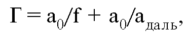
Optical system of the microscope. How to calculate the magnification of a telescope
Ø General principles creating an image.
According to the diffraction theory of Abbe imaging, a complete image of an object, reproduced using a microscope, is obtained by superimposing two images that are formed due to the phenomena of diffraction (primary image) and interference (secondary image) of the light flux passing through the object. The principle of operation of the microscope is simple: a beam of light rays is directed by a condenser lens through the sample, and the resulting image is then magnified using lenses.
Consider the principle of creating an image in more detail. The object (preparation) is placed on a glass slide. The condenser concentrates on the object a beam of light reflected from the mirror. The source of light in a microscope is most often a special illuminator; sometimes a mirror directs normal daylight onto an object. Diaphragms - field and aperture limit the light beam and reduce the proportion of scattered light in it that falls on the preparation "from the side" and is not involved in image formation.
The appearance of an image of a specimen in a microscope can be described in its basic (albeit the simplest) terms within the framework of geometric optics. The rays of light emanating from the object, refracted in the lens, create an inverted and enlarged real image - an optical image of the object. This image is viewed through an eyepiece. In visual observation, the microscope is focused so that the optical image is directly behind the front focus of the eyepiece. Under these conditions, the eyepiece works like a magnifying glass: giving additional magnification, it forms a virtual image (still inverted); passing through the optical media of the observer's eye, the rays from the imaginary image create a real image of the object on the retina of the eye. Usually the virtual image is located at the distance of best vision from the eye.
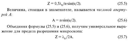
Ø Numerical (numerical) aperture and resolution.
The numerical aperture of the lens (A) is the product of the sine of half the aperture and the refractive index of the medium between the object and the lens: A \u003d n x sin α, where
n is the refractive index of the medium lying between the object of observation and the lens,
α - half the angle of the light beam coming from the point and entering the lens. The numerical aperture defines the series the most important properties microscope: image brightness, "penetrating" and "displaying" abilities, i.e. the degree of similarity of the image with the subject. The larger the numerical aperture, the finer details the lens can reproduce.
Resolution is the ability of the eye or optical instrument to distinguish the smallest distance between images of two adjacent points (lines) that differ as two individual images. In other words, if we bring two points that are distant from each other closer, then upon reaching a certain critical distance, they will merge and will be perceived as one. Resolution (resolution0 is the smallest distance at which two nearby points of an object are still perceived separately.
For example, the naked human eye has a resolution of about 1/10 mm, or 100 microns. This means that if a person looks at two lines that are less than 100 microns apart, they merge into one.
d = (0.61λ)/(nxsinα)
Thus, the resolution depends on the wavelength of light, the refractive index of the medium. In addition, the resolution has a limit due to the wave properties of light. According to general pattern, observing an object in any radiation with a wavelength l, it is impossible to distinguish the elements of the object separated by distances much smaller than l. This pattern also manifests itself in a microscope, and its quantitative expression is somewhat different for self-luminous and non-luminous objects.
Ø Microscope magnification.
The magnification of a microscope should be understood as the ratio of the size of the image of the drug on the retina, formed when viewed through a microscope, to the size of the same drug, obtained on the retina when viewed with the naked eye. The overall magnification of a microscope is the product of the magnification of the objective and the magnification of the eyepiece. If one or more magnifying systems are located between them, then the total magnification of the microscope is equal to the product of the magnifications of all optical systems, including intermediate ones: objective, eyepiece, binocular attachment, wholesaler or projection systems: Г m = o6 x Г ok x ql x q2 x ... , where GM is the total magnification of the microscope; about - lens magnification; Gok - magnification of the eyepiece; ql, q2 ... - increase in additional systems. For example, in domestic microscopes BIOLAM R-11, S-11, the monocular attachment has no magnification, therefore, the total magnification of the microscope with a 90 x objective and 10 x eyepiece will be: 90 x 10 = 900 x R-15, BIOLAM I, has its own magnification of 1.5x. Therefore, the total magnification of the microscope in this case will be: 90x10x1.5 = 1350x. The magnification of the microscope can reach 2000x.
Ø Useful magnification of a microscope.
Useful magnification is the apparent magnification at which the observer's eye will fully utilize the resolving power of the microscope, that is, the resolving power of the microscope will be the same as the resolving power of the eye. The useful magnification of the microscope should be no more than 1000 numerical apertures of the objective and no less than 500:500A rev.<Г м < 1000 А об, где Аоб - числовая апертура объектива. Например, для объектива 90x1,25 полезное увеличение микроскопа лежит в диапазоне 625х-1250х. При большем увеличении изображение становится нечетким и малоконтрастным, с пониженной разрешающей способностью; при меньшем увеличении - изображение объекта, несмотря на четкость и повышенный контраст, становится настолько мелким, что элементы объекта практически неразличимы.
An example of calculating the useful magnification and selecting optics if it is necessary to select an eyepiece. Lens 90x1.25 MI; binocular attachment AU-12, having its own magnification of 1.5x, the numerical aperture of the objective - A o6 = = 1.25.
The lower limit of the magnification of the microscope should be: 500x1.25 = 625.
The upper limit of the magnification of the microscope should be: 1000x1.25= 1250.
Total magnification of the lens and binocular attachment: 90x 1.5 = 135.
Thus, the minimum magnification of the eyepiece will be: 625: 135 = 4.6x, and the maximum magnification - 1250: 135 = 9.2 x.
Thus, objects with a size of at least 0.2 - 0.3 microns are resolved in an optical microscope. In order for these objects to be visible to the eye as well, magnification K m microscope should not be less than the value determined by the ratio of the resolution limits Z eyes and microscope Z m : K m \u003d Z hl / Z m , substituting into this formula the value Z , we get K m = 2A Z ch / l .
Z eyes(at the distance of best vision) in the range 2 - 4 is from 140 to 280 microns. Substituting them, as well as l = 0.555 µm, into the formula, we find the range of values for the useful magnification of the microscope: 500A< K м < 1000А . These increases are called useful, because. with them, the eye distinguishes all elements of the structure of the object that are resolvable under a microscope. Substituting the numerical aperture of the oil immersion system (n = 1.43) into the formula, we obtain the following inequality for the useful magnifications of such a microscope: 700 < K м < 1400 .
Special microscopy techniques:
measuring the size of small objects,
microprojection, microphotography,
phase contrast method,
dark field method (ultramicroscopy).
1. Measuring Small Objects .
Determining the size of a microscopic object is done using scale scales applied to a glass plate, called ocular and objective micrometers.
An ocular micrometer is placed between the lenses of the eyepiece so that its scale is in the plane of the intermediate image formed by the lens. At the same time, the image of the scale is observed in the eyepiece, combined with the image of the microscoped object. Given the division value of the micrometer scale, you can determine the size of this image given by the lens, and by dividing the obtained data by the known magnification of the lens K about - the actual dimensions of the object.
If the division of the ocular micrometer is not known, then it can be determined using an objective micrometer with a known division (usually 0.01 mm). An object micrometer is placed in place of the preparation and a combined image of both scales is observed in the eyepiece.
2. Microprojection and microphotography .
The imaginary nature of the image in the microscope is due to the fact that the intermediate real image formed by the objective is located closer to the front focus of the eyepiece. If this condition is violated, for example, by turning the eyepiece so that the image produced by the lens is farther than the focal length of the eyepiece, then the latter will give a real image that can be projected onto a screen or film. The method of observing the actual image of an object on the screen is called microprojection. Usually, the microscope is placed horizontally and the object is illuminated with a strong light source.
Photographing the actual image obtained in this way is called microphotography. Usually, a special photo attachment to the microscope is used, which is a camera that is put on the ocular end of the microscope tube.
3. Phase contrast method .
The method is used to observe low-contrast objects; it is based on the use of the phase difference, which is formed when light passes through various structures of the object under study.
4. Dark field method (ultramicroscopy).
It is a method of microscopy of unfixed and unstained objects. Observation of such objects in transmitted light does not give the desired results due to the lack of contrast between the elements of the structure of the object, as well as between the object and the environment. In these cases, the dark field observation method is used, which is carried out by using a special condenser in a conventional biological microscope.
The observer's eye will be able to perceive two points as separate if the angular distance between them is not less than the angular limit of the eye's resolution. In order for the observer's eye to make full use of the resolution of the microscope, it is necessary to have an appropriate apparent magnification.
Useful magnification is the apparent magnification at which the observer's eye will fully utilize the resolving power of the microscope, i.e. the resolving power of the microscope will be the same as the resolving power of the eye.
If two points in the front focal plane of the microscope are located at a distance 𝜎 from each other, then the angular distance between the images of these points. From expressions (6.11) and (6.8) one can derive the apparent magnification of the microscope:
Since the typical exit pupil of a microscope is about 0.5-1 mm, the angular resolution limit of the eye is 2" - 4". If we take the average wavelength in the visible region of the spectrum (0.5 μm), then for a useful magnification of the microscope, we can derive the dependence:
A microscope with a visible magnification of less than 500A does not allow the eye to distinguish all the subtleties of the structure of the object, which are depicted as separate by this lens (<). Использование видимого увеличения больше 1000А нецелесообразно, так как разрешающая способность объектива не позволяет полностью использовать разрешающую способность глаза (>).
End of work -
This topic belongs to:
Introduction to the specialty
Federal State Budgetary Educational Institution... of Higher Professional Education... Tula State University...
If you need additional material on this topic, or you did not find what you were looking for, we recommend using the search in our database of works:
What will we do with the received material:
If this material turned out to be useful for you, you can save it to your page on social networks:
| tweet |
All topics in this section:
By discipline
"Introduction to the specialty" Direction of training: 200400 "Optical engineering" Training profile: "Optico-electronic
The history of the development of optics.
Optics is the study of the nature of light, light phenomena and the interaction of light with matter. And almost all of its history is the history of the search for an answer: what is light? One of the first theories of light - theo
Area of professional activity
The field of professional activity of a bachelor in the direction of training 200400 with a training profile "Optico-electronic devices and systems" is research, development, preparation and
The main courses of lectures are a brief description. Organization of training.
Basic courses of lectures. Mechanics Materials Science and Technology of Structural Materials
The structure of the eye
Figure 2.1. a section of the eyeball is shown and the main details of the eye are shown. The eye is a spherical body (eyeball), almost completely covered with opaque
Accommodation
Accommodation is the ability of the eye to adapt to a clear distinction between objects located at different distances from the eye. Accommodation occurs by changing the curvature of the surface
The structure of the retina
The retina is a complex interweaving of nerve cells and nerve fibers that connect nerve cells to each other and connect the eye to the cerebral cortex. Main photosensitive elements
Spectral sensitivity
Optical devices that work in conjunction with the eye deal with that part of the radiation flux that affects the eye. It includes the visible region of the spectrum in the wavelength range 380 - 780 nm
Adaptation
Adaptation of the eye to changing light conditions is called adaptation. Distinguish between dark and light adaptation. Dark adaptation occurs during the transition from large bright
Field of view of the eye
The total field of view of the eye is enormous, larger than that of any other optical device (125° vertically and 150° horizontally), but in reality, for a clear distinction,
Limit of eye resolution
In any optical system, there is a finite limit to the clarity of detail. For optics designers, of great interest is the value of the lower limit of the resolution of the eye of two adjacent t
Visual defects and their correction
If the far point of the eye is infinitely removed, then such an eye is called normal or emmetropic. At the same time, the eye distinguishes objects well both far and near. This means that the optical apparatus
Myopia
There are two reasons for myopia. The first is an elongated eyeball with normal refractive power of the eye. Another reason is too high optical power of the optical system of the eye (more than 60 di
farsightedness
Farsightedness is caused by a weak optical power of the optical system of the eye for a given length of the eyeball (either a short eye with normal optical power, or a low optical power of the eye with
Astigmatism
The cause of astigmatism lies either in an irregular, non-spherical shape of the cornea (in different sections of the eye passing through the axis, the radii of curvature are not the same), or in non-centric with respect to the optical
Optical system
Optical system - a set of optical media separated by optical surfaces and containing diaphragms. The optical system is designed to form an image by means of redistribution
Characteristics of the subject and image
An object is a set of points from which rays come out that enter the optical system. All possible set of points forms the space of objects. The optical system divides all p
Pupillary characteristics
Not all the rays that come from the object pass through the optical system. Limiting the size of beams of rays is the result of the joint action of all apertures available in the optical system. However, mo
Spectral characteristics
Spectral characteristics are necessary to match the range of wavelengths that an object emits and in which an image is formed. Usually, all calculations of the path of rays in an optical system are done for
Transfer characteristics
Transfer characteristics show how the device converts an object into an image. The effect of the optical system on the radiation emanating from the object is reduced primarily to the transformation
Scale transfer characteristics
Scale transfer characteristics describe the transmission of the size and shape of an object by an optical system, that is, the transformation of coordinates on the object into coordinates on the image. Generalized
Energy transfer characteristics
Energy transfer characteristics describe the transfer of the energy of the object by the device. Since not all rays emanating from the object pass through the optical system, and since in the optical system itself
Structural transfer characteristics
Imaging devices with the same magnification and luminosity can produce images of different quality in terms of conveying the fine structure of the object (more or less sharp, with more or less
Cameras
The camera is perhaps the most common optical parting. Almost everyone has a camera these days. Moreover, modern compact cameras are so easy to use.
Relative aperture of a photographic lens
The relative aperture is the absolute value of the ratio of the diameter of the aperture stop to the rear focal length of the lens: (4.3) Since the value calculated
Depth of field of a photographic lens
Since all lenses have aberrations, one point of the object will always appear as a circle of confusion. However, when viewing the image with the eye, this is not noticed, since the resolution
Wide angle (short throw)
For wide angle lenses focal length less than the diagonal of the frame. Wide-angle lenses are characterized by a short focal length in the range of approximately mm. The field of view of such
Narrow-angle (long-focus)
Narrow-angle lenses have a focal length greater than the diagonal of the frame, and a field of view of less than 40°. The focal length of such lenses is more than 50 mm. Usually as long-focus are used about
Zoom Lenses
Lenses with variable focal length (ZOOM-lenses) allow you to obtain images of various scales at a constant distance to the subject. For example, using a lens with a range
Focus systems
In creating high-quality images, one of the most important functions is performed by the camera's focusing system, that is, the focusing process. In the simplest cameras, it is not carried out
exposition
Exposure is the amount of light that hits a photo:< экспозиция > = < интенсивность света > ∙ < время воздействия >. Light intensity control
Features of digital cameras
AT recent times digital cameras are becoming more and more popular. Unlike film, digital cameras The image receiver is a CCD (Charge Coupled Device)
Telescopic system
Telescopic system - an optical system with which you can view an enlarged image of a distant object. These instruments include binoculars, spotting scopes,
Visible magnification of the telescopic system
The apparent magnification of a telescopic system can be expressed in terms of the ratio of the focal length of the objective to the focal length of the eyepiece: If the apparent magnification is positive
Diameters of the entrance and exit pupils of the telescopic system
Exit pupil diameter is determined by the pupil of the eye: . (5.3) When observing objects through a telescopic instrument, the eye must be located in the plane of the exit pupil, then the weight
Visible loupe magnification
According to the definition, the apparent magnification of a magnifying glass is calculated as the ratio of the tangent of the angle at which an object is seen through a magnifying glass to the tangent of the angle at which an object is observed with the naked eye with
Magnifying glass field of view
On fig. 6.3 shows a magnifying glass with a diameter Dn. The pupil of the observer's eye with a diameter of Dra is located at a distance S "from the magnifying glass. The size of the field 2a" in the image space is determined by
Microscope
The microscope is designed to observe small objects with a high magnification and with a higher resolution than a magnifying glass gives. The optical system of a microscope consists of two parts: an objective and a lens.
Microscope magnification
The action of a microlens is characterized by its linear increase: where is the focal length of the microlens, Δ is the distance between the rear focus of the lens and the re
Microscope resolution
One of the most important characteristics microscope is its resolving power. According to Abbe's diffraction theory, the linear limit of microscope resolution, that is, the minimum distance between points
Observation methods
Usually, objects examined under a microscope do not themselves glow and, therefore, need extraneous illumination. In many cases, the objects in question are thin slices of transparent
bright field method
The bright field method in transmitted light is used in the study of transparent preparations in which different parts of the structure absorb light differently (thin stained sections of animals and growing
Dark field method
The dark field method in transmitted light is used in biology, colloid chemistry, mineralogy and other areas to obtain images that are transparent, non-absorbing, and therefore not visible during observation.
Research method in polarized beams
The research method in polarized beams is used in transmitted and reflected light for the so-called anisotropic objects with birefringence or reflection. Such objects
Phase contrast method
The phase contrast method makes it possible to obtain contrast images transparent and colorless objects. Such objects include, for example, unstained biological preparations, non-herbal
Light microscopes
Biological microscopes (MULTISCOPE™, LABOROSCOPE™, INVERTOSCOPE™ series produced by LOMO) are the most universal and therefore the most widespread. Modern biological micro
Electron microscopes
Electron microscope built on the same principle of image acquisition as optical, but instead of visible light it uses an electron beam. The role of lenses in the electron microscope
Scanning microscopes
Scanning microscopes are based on a different imaging principle that overcomes the diffraction limit of resolution. The principle of operation of such microscopes is based on scanning
Lighting systems
A lighting system is a device designed to illuminate non-luminous objects. In most cases, it is impossible to provide the required illumination of the object and its uniform
Condenser
If the illuminated object is at a finite distance, then a condenser is used to illuminate it. There are two options for the optical scheme of the condenser. In the first scheme, the optical system pr
Lighting optical systems
Lighting optical systems can improve the quality of lighting, use most light source and provide more uniform illumination of the object. Main elements
Searchlight
A searchlight is an optical system that concentrates the luminous flux of a light source into a narrow beam to illuminate distant objects or to transmit signals to long distances(Fig. 7.7).
Lighting systems for projection devices
Projection devices are designed to obtain images of objects of the required scale on the screen. The main devices of the projector are lighting, providing a uniform and intense
Illumination systems of microscopes
Since most objects examined with a microscope are not self-luminous, additional light sources are required to work with them. The lighting system of the microscope should provide
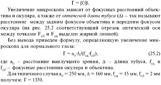
2. Optical system of the microscope.
3. Magnification of the microscope.
4. Permission limit. Resolution of the microscope.
5. Useful magnification of the microscope.
6. Special techniques of microscopy.
7. Basic concepts and formulas.
8. Tasks.
The ability of the eye to distinguish fine details of an object depends on the size of the image on the retina or on the angle of view. To increase the angle of view, special optical devices are used.
25.1. magnifying glass
the simplest optical instrument to increase the angle of view is a magnifying glass, which is a short-focus converging lens (f = 1-10 cm).
The object in question is placed between the magnifying glass and its front focus in such a way that its imaginary image is within the accommodation for a given eye. Usually, planes of far or near accommodation are used. The latter case is preferable, since the eye does not get tired (the annular muscle is not tense).
Let's compare the angles of view at which the object is seen, considered by the "naked" normal eye and with a magnifying glass. The calculations are performed for the case when the imaginary image of the object is obtained at infinity (the far limit of accommodation).
When examining an object with the naked eye (Fig. 25.1, a), to obtain the maximum angle of view, the object must be placed at the distance of best vision a 0. The angle of view, under which the object is seen in this case, is equal to β \u003d B / a 0 (B is the size of the object).
When examining an object with a magnifying glass (Fig. 25.1, b), it is placed in the front focal plane of the magnifying glass. In this case, the eye sees an imaginary image of the object B "located in an infinitely distant plane. The angle of view under which the image is seen is equal to β" ≈ V / f.
Rice. 25.1. Viewing angles: a- to the naked eye; b- using a magnifying glass: f - focal length of the magnifying glass; N - nodal point of the eye
Magnifying glass- angle of view ratioβ", under which you can see the image of the object in a magnifying glass, to the angle of viewβ, under which the object is visible to the "naked" normal eye from the distance of best sight:
The magnification of a magnifying glass for a near-sighted and far-sighted eye is different, since they have different distances of best vision.
We give without derivation the formula for the magnification given by a magnifying glass used by a near-sighted or far-sighted eye when forming an image in the plane of far accommodation:
 where a is the far limit of accommodation.
Formula (25.1) suggests that by reducing the focal length of the magnifying glass, an arbitrarily large increase can be achieved. Basically it is. However, when reducing the focal length of the magnifier and maintaining its size, such aberrations arise that nullify the entire effect of magnification. Therefore, single lens magnifiers usually have 5-7x magnification.
To reduce aberrations, complex magnifiers are made, consisting of two or three lenses. In this case, it is possible to achieve a 50-fold increase.
25.2. Optical system of the microscope
Greater magnification can be achieved by examining with a magnifying glass the actual image of the object created by another lens or lens system. Such an optical device is implemented in a microscope. The magnifying glass in this case is called eyepiece, and the other lens lens. The path of the rays in the microscope is shown in Fig. 25.2.
Object B is placed near the front focus of the lens (F rev) so that its actual, enlarged image B "is between the eyepiece and its front focus. When
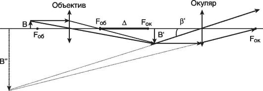 Rice. 25.2. The path of rays in a microscope.
In this case, the eyepiece gives a virtual magnified image B", which is examined by the eye.
By changing the distance between the object and the lens, the image B "is in the plane of far accommodation of the eye (in this case, the eye does not get tired). For a person with normal vision, B" is located in the focal plane of the eyepiece, and B "is obtained at infinity.
25.3. Microscope magnification
The main characteristic of a microscope is its angular increase. This concept is analogous to the angular magnification of a magnifying glass.
Microscope magnification- angle of view ratioβ", under which you can see the image of the object in eyepiece, to the angle of viewβ, under which the object is visible to the "naked" eye from the distance of best vision (a 0):
 25.4. Permission limit. Microscope resolution
One may get the impression that, by increasing the optical length of the tube, one can achieve an arbitrarily large magnification and, therefore, consider the smallest details of the object.
However, taking into account the wave properties of light shows that the dimensions small parts, distinguishable with a microscope, there are restrictions associated with diffraction light passing through the aperture of the lens. Due to diffraction, the image of the illuminated point is not a point, but small light circle. If the considered details (points) of the object are far enough away, then the lens will give their images in the form of two separate circles and they can be distinguished (Fig. 25.3, a). The smallest distance between distinguishable points corresponds to the "touch" of the circles (Fig. 25.3, b). If the points are very close, then the "circles" corresponding to them overlap and are perceived as one object (Fig. 25.3, c).
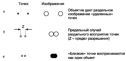 Rice. 25.3. Resolution
The main characteristic showing the capabilities of the microscope in this regard is permission limit.
Resolution Limit microscope (Z) - the smallest distance between two points of an object at which they are distinguishable as separate objects (i.e. perceived in a microscope as two points).
The reciprocal of the resolution limit is called resolving power. The smaller the resolution limit, the greater the resolution.
The theoretical limit of a microscope's resolution depends on the wavelength of the light used for illumination and on angular aperture lens.
Angular aperture(u) - the angle between the extreme rays of the light beam entering the objective lens from the object.
 Let us indicate without derivation the formula for the resolution limit of a microscope in air:
where λ is the wavelength of the light that illuminates the object.
Modern microscopes have an angular aperture of up to 140°. If accept λ = 0.555 µm, then we get the value Z = 0.3 µm for the resolution limit.
25.5. Useful microscope magnification
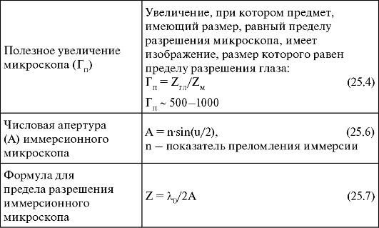
Let us find out how large the magnification of the microscope should be for a given limit of resolution of its objective. Let us take into account that the eye has its own limit of resolution due to the structure of the retina. In Lecture 24 we got next estimate for eye resolution limit: Z GL = 145-290 µm. In order for the eye to be able to distinguish the same points that the microscope separates, magnification is necessary.
This increase is called useful increase.
Note that when using a microscope to photograph an object in formula (25.4), instead of Z GL, one should use the film resolution limit Z PL.
Useful microscope magnification- magnification at which an object having a size equal to the resolution limit of a microscope has an image whose size is equal to the resolution limit of the eye.
Using the estimate obtained above for the resolution limit of the microscope Z m ≈0.3 μm), we find: G p ~ 500-1000.
It does not make sense to achieve a larger value for magnifying the microscope, since it will not be possible to see any additional details anyway.
Useful microscope magnification - this is a reasonable combination of resolving powers of both the microscope and the eye.
25.6. Special Microscopy Techniques
Special microscopy techniques are used to increase the resolution (reduce the resolution limit) of a microscope.
1. Immersion. In some microscopes, to reduce resolution limit the space between the lens and the object is filled with a special liquid - immersion. Such a microscope is called immersion. The effect of immersion is to reduce the wavelength: λ = λ 0 /n, where λ 0 - the wavelength of light in vacuum, and n is the immersion refractive index. In this case, the resolution limit of the microscope is determined by the following formula (generalization of formula (25.3)):
 Note that special lenses are created for immersion microscopes, since the focal length of the lens changes in a liquid medium.
2. UV microscopy. For decreasing resolution limit use shortwave ultraviolet radiation, invisible to the eye. In ultraviolet microscopes, a micro-object is examined in UV rays (in this case, the lenses are made of quartz glass, and registration is carried out on photographic film or on a special luminescent screen).
3. Measurement of the size of microscopic objects. Using a microscope, you can determine the size of the observed object. To do this, use an ocular micrometer. The simplest ocular micrometer is a round glass plate on which a scale with divisions is applied. The micrometer is set in the plane of the image received from the lens. When viewed through the eyepiece, the images of the object and the scale merge, it is possible to count out what distance on the scale corresponds to the measured value. Preliminarily determine the value of division of the ocular micrometer according to the known object.
4. Microprojection and microphotography. Using a microscope, you can not only observe an object through the eyepiece, but also photograph it or project it onto a screen. In this case, special eyepieces are used, which project the intermediate image A "B" onto the film or onto the screen.
5. Ultramicroscopy. The microscope allows you to detect particles whose sizes lie outside its resolution. This method uses oblique illumination, due to which the microparticles are visible as bright dots on a dark background, while the structure of the particles cannot be seen, only the fact of their presence can be established.
The theory shows that no matter how powerful the microscope is, any object smaller than 3 microns in size will be represented in it simply as a single point, without any details. But this does not mean that such particles cannot be seen, monitored, or counted.
To observe particles whose dimensions are less than the resolution limit of a microscope, a device called ultramicroscope. The main part of the ultramicroscope is a strong illuminating device; particles illuminated in this way are observed in an ordinary microscope. Ultramicroscopy is based on the fact that small particles, suspended in a liquid or gas, become visible in strong side illumination (recall the dust particles visible in a sunbeam).
25.8. Basic concepts and formulas
 End of table
 25.8. Tasks
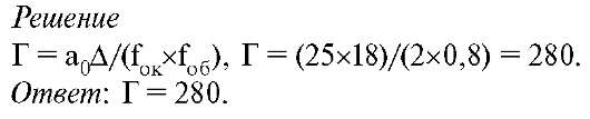
1. A lens with a focal length of 0.8 cm is used as a microscope objective with an eyepiece focal length of 2 cm. The optical length of the tube is 18 cm. What is the magnification of the microscope?
 2.
Determine the resolution limit of dry and immersion (n = 1.55) lenses with an angular aperture u = 140 o. Take the wavelength equal to 0.555 µm.
2.
Determine the resolution limit of dry and immersion (n = 1.55) lenses with an angular aperture u = 140 o. Take the wavelength equal to 0.555 µm.
 3.
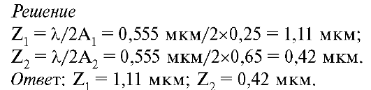
What is the limit of resolution at a wavelength λ
\u003d 0.555 microns, if the numerical aperture is: A 1 \u003d 0.25, A 2 \u003d 0.65?
3.
What is the limit of resolution at a wavelength λ
\u003d 0.555 microns, if the numerical aperture is: A 1 \u003d 0.25, A 2 \u003d 0.65?
 4.
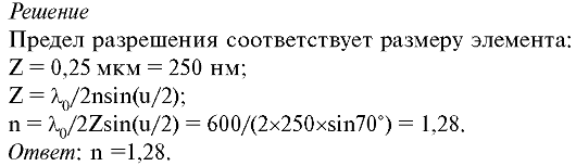
With what refractive index should the immersion liquid be taken in order to examine a subcellular element with a diameter of 0.25 μm in a microscope when viewed through an orange light filter (wavelength 600 nm)? The aperture angle of the microscope is 70°.
4.
With what refractive index should the immersion liquid be taken in order to examine a subcellular element with a diameter of 0.25 μm in a microscope when viewed through an orange light filter (wavelength 600 nm)? The aperture angle of the microscope is 70°.
 5.
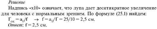
On the rim of the magnifier there is an inscription "x10" Determine the focal length of this magnifier.
5.
On the rim of the magnifier there is an inscription "x10" Determine the focal length of this magnifier.
 6.
Focal length of the microscope lens f 1 = 0.3 cm, tube length Δ
\u003d 15 cm, magnification G \u003d 2500. Find the focal length F 2 of the eyepiece. Best vision distance a 0 = 25 cm.
6.
Focal length of the microscope lens f 1 = 0.3 cm, tube length Δ
\u003d 15 cm, magnification G \u003d 2500. Find the focal length F 2 of the eyepiece. Best vision distance a 0 = 25 cm.
The microscope is designed to observe small objects with a high magnification and with a higher resolution than a magnifying glass gives. The optical system of a microscope consists of two parts: an objective and an eyepiece. The microscope objective forms a true magnified inverse image of the object in the front focal plane of the eyepiece. The eyepiece acts like a magnifying glass and forms a virtual image at the best viewing distance. In relation to the entire microscope, the object under consideration is located in the front focal plane.
Microscope magnification
The action of a microlens is characterized by its linear increase: V about \u003d -Δ / F \ "about * F \" about - the focal length of the microlens * Δ - the distance between the rear focus of the lens and the front focus of the eyepiece, called the optical interval or the optical length of the tube.
The image created by a microscope objective at the front focal plane of the eyepiece is viewed through the eyepiece, which acts like a loupe with apparent magnification:
G ok =¼ F ok
The total magnification of the microscope is defined as the product of the objective magnification and the magnification of the eyepiece: G=V rev *G ok
If the focal length of the entire microscope is known, then its apparent magnification can be determined in the same way as for a magnifying glass:
As a rule, the magnification of modern microscope objectives is standardized and is a series of numbers: 10, 20, 40, 60, 90, 100 times. Eyepiece magnifications also have quite definite values, for example, 10, 20, 30 times. All modern microscopes have a set of objectives and eyepieces that are specially designed and manufactured to fit together so they can be combined to obtain different magnifications.
Microscope field of view
The field of view of the microscope depends on the angular field of the eyepiece ω , within which an image of sufficiently good quality is obtained: 2y=500*tg(ω)/G * G - microscope magnification
For a given angular field of the eyepiece, the linear field of the microscope in the space of objects is the smaller, the greater its apparent magnification.
Microscope Exit Pupil Diameter
The microscope exit pupil diameter is calculated as follows:
where A is the front aperture of the microscope.
The exit pupil of a microscope is usually slightly smaller than the pupil of the eye (0.5–1 mm).
When observing through a microscope, the pupil of the eye must be aligned with the exit pupil of the microscope.
Microscope resolution
One of the most important characteristics of a microscope is its resolution. According to Abbe's diffraction theory, the linear resolution limit of a microscope, that is, the minimum distance between the points of an object that are depicted as separate, depends on the wavelength and the numerical aperture of the microscope:
Maximum achievable resolution optical microscope can be calculated from the expression for the microscope aperture . If we take into account that the maximum possible value of the sine of the angle is unity, then for the average wavelength we can calculate the resolution of the microscope: ![]()
There are two ways to increase the resolution of the microscope: * By increasing the aperture of the objective, * By decreasing the wavelength of light.
Immersion
In order to increase the lens aperture, the space between the object under consideration and the lens is filled with the so-called immersion liquid - a transparent substance with a refractive index greater than one. Water, cedar oil, glycerin solution and other substances are used as such a liquid. The apertures of high-magnification immersion objectives reach the value , then the maximum achievable resolution of an immersion optical microscope will be. ![]()
The use of ultraviolet rays
To increase the resolution of the microscope in the second way, ultraviolet rays are used, the wavelength of which is shorter than that of visible rays. In this case, special optics that are transparent to ultraviolet light must be used. Since the human eye does not perceive ultraviolet radiation, it is necessary either to resort to means that convert an invisible ultraviolet image into a visible one, or to photograph the image in ultraviolet rays. At a wavelength, the resolution of the microscope will be. ![]()
In addition to improving the resolution, the method of observation in ultraviolet light has other advantages. Usually, living objects are transparent in the visible region of the spectrum, and therefore they are preliminarily stained before observation. But some objects nucleic acids, proteins) have selective absorption in the ultraviolet region of the spectrum, due to which they can be "visible" in ultraviolet light without staining.


, poultry farming")


- Burns, Robert - short biography
- The concept of common vocabulary and vocabulary of limited use
- Nancy Drew: The Captive Curse Walkthrough Nancy Drew Curse of Blackmoore Manor Walkthrough
- Deadpool - Troubleshooting
- Won't start How to Survive?
- What to do if bioshock infinite won't start
- Walkthrough Nancy Drew: Alibi in Ashes
- Spec Ops: The Line - game review, review Spec ops the line crashes on missions
- Room escape level 1 walkthrough
- Processing tomatoes with boric acid How much will 2 grams of boric acid
- Cucumber Grass (Borago)
- Bioinsecticide Lepidocid: purpose, properties and application procedure Lepidocide waiting period
- How to change the language to Russian in steam
- Dendrobium noble: room care
- Morphology of plants general concepts - document
- Planting, propagation and care of bamboo at home, photo Growing bamboo from seeds
- How to strengthen the cellular signal for the Internet in the country
- Sanskrit reveals the forgotten meaning of Russian words (2 photos)
- The oldest language Sanskrit programming language of the future Dead language Sanskrit
- Who has dominion over all the earth?