How to draw a human head? Proportions of the head and face. Brain: structure and functions, general description
Neck bones consist of seven cervical vertebrae, which include the upper part of the spine. Large muscles that influence the shape of the neck are the trapezius muscle in the back and the sternomastoideus muscle in the front. They run from the back of the ear down to the inner ends of the clavicles.
Neck able to move in all directions: Tilt your head back and forth, sideways to either shoulder, and rotate it from side to side 180 degrees.
Apart from the sound-conducting structures in the ears, only the jaw moves from the joints of the head. All other bones of the skull are rigidly linked and immovable.
The muscles of the face can be conditionally divided into two types:
The proportions of the human head. Simple construction method
The face is, of course, the most expressive part of the body. Here simple construction method which will help you see which simple forms head consists. The average proportions of a human head are shown in the figure below. The height of the head is about the same as its width when viewed from the side, so in profile it fits into a square. Seen from the front, the width of the head is much less than its height.. 
Start by drawing the head in profile. Draw a circle for the skull and then add two lines on the front of the face to represent the jaw as shown in the image above. 
The most common rookie mistake when depicting the head is that head looks flat. Therefore, as soon as possible, try to draw three-quarter view of the head, trying to display the roundness and solidity of the forms. Use light reference lines to indicate the center line of the face and the position of the eyes.  The figure shows a rather spineless face, but this stage it is important to understand the basic forms. The faces of people differ from the average sample in many respects, exactly how will be discussed in detail in next lessons. These drawings represent the first step. If you draw hundreds of these simplified heads, you will come to understand all the subtleties of the relief. human face and be able to fill your drawings with life and character.
The most important thing to understand is that the face is not just a flat surface with details superimposed on it. To draw faces successfully, you must understand the 3D shape of the surface - that's why we started with a featureless, average face that can belong to either a man or a woman. 
If you look at the hazy newspaper photo of the crowd below, you will see that each face differs from the rest only in the display of light and shadow on it, and not in the shape of the eyes or lips. If you can imagine face as a template, in this case you will avoid the common mistake of creating lifeless mechanisms of eyes, noses and mouths, indistinguishable from each other.  That's why practice drawing the face template with the addition of light and shadow until you fully understand its shape.
  The images above show several basic forms faces and heads at different angles. Once again, it is important now that you understand the real shape of the surface and not just a plan.
The article used materials from the book Ron Tiner "Figure Drawing without a model".
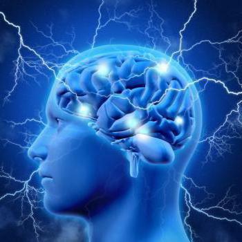
The brain is the main regulator of the functions of any living organism, one of the elements Until now, medical scientists have been studying the features of the brain and discovering new incredible possibilities. This is a very complex organ that connects our body with the external environment. The parts of the brain and their functions regulate all life processes. External receptors catch signals and inform any part of the brain about incoming stimuli (light, sound, tactile, and many others). The response is immediate. Let's take a closer look at how our head "processor" works.
General description of the brain
The parts of the brain and their functions completely control our life processes. The human brain consists of 25 billion neurons. This incredible number of cells forms the gray matter. The brain covers several layers:
- soft;
- hard;
- arachnoid (cerebrospinal fluid circulates here).
Liquor is a cerebrospinal fluid, in the brain it plays the role of a shock absorber, a protector from any impact force.
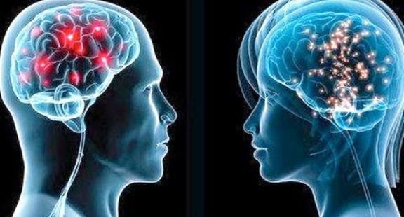
In both men and women, the brain is developed in exactly the same way, although its weight is different. More recently, the debate has subsided that brain weight plays some role in mental development and intellectual abilities. The conclusion is unambiguous - it is not. The weight of the brain is approximately 2% of the total mass of a person. In men, its average weight is 1,370 g, and in women - 1,240 g. The functions of the parts of the human brain are developed in a standard way, vital activity depends on them. Mental abilities depend on the quantitative connections created in the brain. Each brain cell is a neuron that generates and transmits impulses.
The cavities inside the brain are called ventricles. The cranial paired nerves go to different departments.
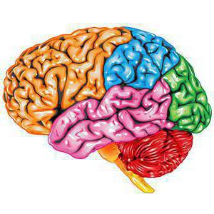
Functions of the brain regions (table)
Every part of the brain has a job to do. The table below clearly demonstrates this. The brain, like a computer, clearly performs its tasks, receiving commands from the outside world.
The functions of the brain regions, the table reveals schematically and succinctly.

Let's look at the brain regions in more detail below.
Structure
The picture shows how the brain works. Despite this, the most significant part is occupied by all parts of the brain and their functions play a huge role in the functioning of the body. There are five main divisions:
- final (of the total mass is 80%);
- posterior (bridge and cerebellum);
- intermediate;
- oblong;
- average.
At the same time, the brain is divided into three main parts: the brain stem, the cerebellum, and the two cerebral hemispheres.

telencephalon
It is impossible to briefly describe the structure of the brain. To understand the parts of the brain and their functions, it is necessary to study their structure closely.
The telencephalon stretches from the frontal to the occipital bone. Two cerebral hemispheres are considered here: left and right. This section is different from others. largest number furrows and convolutions. The development and structure of the brain are closely linked. Experts have identified three types of bark:
- ancient (with olfactory tubercle, anterior perforated substance, semilunar subcallosal and lateral subcallosal gyrus);
- old (with dentate gyrus - fascia and hippocampus);
- new (represents the rest of the cortex).
The hemispheres are separated by a longitudinal groove, in its depths there is a vault and a corpus callosum, which connect the hemispheres. The corpus callosum itself is lined and belongs to the neocortex. The structure of the hemispheres is quite complex and resembles a multi-level system. Here, the frontal, temporal, parietal and occipital lobes, subcortex and cortex are distinguished. performed by the large hemispheres great amount functions. It is worth noting that the left hemisphere commands the right side of the body, and the right, on the contrary, the left.
Bark
The surface layer of the brain is the cortex, it has a thickness of 3 mm, covers the hemispheres. The structure is made up of vertical nerve cells having processes. The cortex also contains efferent and afferent nerve fibers, as well as neuroglia. The parts of the brain and their functions are discussed in the table, but what is the cortex? Its complex structure has horizontal layering. The building has six layers:
- external pyramidal;
- external granular;
- internal granular;
- molecular;
- internal pyramidal;
- with spindle cells.
Each has a different width, density, shape of neurons. Vertical bundles of nerve fibers give the cortex a vertical striation. The area of the cortex is approximately 2,200 square centimeters, the number of neurons here reaches ten billion.

Parts of the brain and their functions: cortex
The cortex controls several specific bodily functions. Each share is responsible for its own parameters. Let's take a closer look at the functions associated with hotels:
- temporal - controls the sense of smell and hearing;
- parietal - responsible for taste and touch;
- occipital - vision;
- frontal - complex thinking, movement and speech.
Each neuron contacts other neurons, there are up to ten thousand contacts (gray matter). Nerve fibers are white matter. Some part unites the hemispheres of the brain. White matter includes three types of fibers:
- association links connect different cortical areas in one hemisphere;
- commissural connect the hemispheres to each other;
- projection ones communicate with lower formations, have paths of analyzers.
Considering the structure and functions of the brain, it is necessary to emphasize the role of gray and white matter. The hemispheres inside have (gray matter), their main function is the transmission of information. The white matter is located between the cerebral cortex and the basal ganglia. There are four parts here:
- between furrows in convolutions;
- in the outer places of the hemispheres;
- included in the inner capsule;
- located in the corpus callosum.
The white matter located here is formed by nerve fibers and connects the cortex of the convolutions with the underlying sections. form the subcortex of the brain.
The telencephalon - manages all the vital functions of the body, as well as the intellectual abilities of a person.
diencephalon
The brain regions and their functions (table above) include the diencephalon. If you look in more detail, it is worth saying that it consists of ventral and dorsal parts. The hypothalamus belongs to the ventral, and the thalamus, metathalamus, and epithalamus to the dorsal.
The thalamus is a mediator that directs the received irritations to the hemispheres. Often it is referred to as the "optic tubercle". It helps the body quickly adapt to changes in external environment. The thalamus is connected to the cerebellum via the limbic system.
The hypothalamus controls autonomic functions. Influence comes through nervous system, and, of course, the endocrine glands. Regulates the work of the endocrine glands, controls metabolism. The pituitary gland is located directly below it. regulates body temperature, cardiovascular digestive system. The hypothalamus also controls our eating and drinking behavior, regulates wakefulness and sleep.
Rear
The hindbrain includes the pons located in front and the cerebellum, which is located behind. Studying the structure and functions of the parts of the brain, let's take a closer look at the structure of the bridge: the dorsal surface is covered by the cerebellum, the ventral one is represented by a fibrous structure. The fibers are directed transversely in this section. On each side of the bridge, they depart to the cerebellar middle peduncle. In appearance, the bridge resembles a thickened white roller located above the medulla oblongata. The nerve roots exit into the bulbar pontine groove.
The structure of the posterior bridge: on the frontal section, it is clear that it consists of a section of the anterior (large ventral) and posterior (small dorsal) parts. Between them, the trapezoid body serves as a boundary, the transverse thick fibers of which are considered to be the auditory pathway. Conductor function is completely dependent on the hindbrain.
Cerebellum (small brain)
The table "Department of the brain, structure, functions" indicates that the cerebellum is responsible for the coordination and movement of the body. This department is located behind the bridge. The cerebellum is often referred to as the "small brain". It occupies the posterior cranial fossa, covers the rhomboid. The mass of the cerebellum ranges from 130 to 160 g. Above are the large hemispheres, which are separated by a transverse fissure. The lower part of the cerebellum is adjacent to the medulla oblongata.
Two hemispheres are distinguished here, the lower, upper surface and the worm. The boundary between them is called a horizontal deep slit. A lot of cracks cut the surface of the cerebellum, between them there are thin convolutions (rollers). Between the grooves there are groups of convolutions, divided into lobules, they represent the lobes of the cerebellum (posterior, flocculent-nodular, anterior).
The cerebellum contains both grays, and grays are located on the periphery, forming a cortex with molecular and pear-shaped neurons, and a granular layer. Under the cortex there is a white substance that penetrates into the gyrus. In the white matter there are blotches of gray (its nuclei). In cross section, this ratio is similar to a tree. Those who know the structure of the human brain, the functions of its departments, will easily answer that the cerebellum is the regulator of the coordination of the movements of our body.
Midbrain
The midbrain is located in the region of the anterior pons and goes to the papillary bodies, as well as to the optic tracts. Here clusters of nuclei are distinguished, which are called tubercles of the quadrigemina. The structure and functions of the brain regions (table) indicate that this department is responsible for latent vision, the orienting reflex, gives orientation to reflexes to visual and sound stimuli, and also maintains muscle tone human body.
medulla oblongata: brainstem
The medulla oblongata is a natural extension of the spinal cord. That is why the structure has a lot in common. This becomes especially clear if we examine the white matter in detail. It is represented by short and long nerve fibers. In the form of nuclei, gray matter is represented here. Parts of the brain and their functions (the table is presented above) indicates that the medulla oblongata controls our balance, coordination, regulates metabolism, controls breathing and blood circulation. It is also responsible for such important reflexes of our body as sneezing and coughing, vomiting.
The brain stem is divided into the hindbrain and midbrain. The trunk is called the middle, oblong, bridge and diencephalon. Its structure is descending and ascending paths connecting the trunk with the spinal cord and brain. In this part, control over the heartbeat, breathing, articulate speech is carried out.


The brain is the most important part of the human body, responsible for all processes occurring in it. In addition, this organ is often used in symbolic sense: it denotes reason, the ability to think and create, generate new ideas. Therefore, learning how to draw a brain will be interesting and very useful, especially for a novice artist.
Draw the human brain
The human brain is a very complex structure, consisting of several main parts. Even the smallest violation of each of these parts can lead to serious health problems. In order to have an idea about this organ, let's figure out how to draw a human brain.
First, let's draw the so-called big brain, or rather, its hemisphere - we will depict a side view.

Let's start making convolutions on it - the degree of development of the organism depends on their number.

We will depict them on all surfaces.

Now you need to draw other details. Namely, the cerebellum and trunk. Main function cerebellum - coordination in space, muscle tone and balance.
That's all, we coped with the task.
Drawing the brain step by step
Let's practice more in the image of the brain. We will act step by step - it will be easier to learn how to draw this organ in stages. You can draw with what is convenient - with a pencil, charcoal, pen, marker, felt-tip pen, etc.
Let's start with general outlines. The shape of this organ is peculiar, with a certain bend.

Then we will gradually draw the convolutions, one after the other.

They should be on the entire surface - curved, smooth, without sharp corners.

Then we will draw other parts - the bridge, a trunk and a cerebellum. We talked about the functions of the cerebellum in the previous section, while the bridge is responsible for transmitting information from the spinal cord to the brain. The trunk combines all the structures of the central nervous system.
Brain - top view
Before that, we drew the brain exclusively from the side, but now it's time to look at this organ from a different angle, namely from above. So both his hemispheres will be visible. But on this example it will be very interesting to learn how to draw brains with a pencil. After all, the pencil is the very first tool of the artist.
First, the basic shapes. We draw two connected halves. They are somewhat reminiscent of light or very rounded triangles.

After that, we draw inside a lot of short curved lines - gyrus.

Let's outline the shadow parts around the edges. At first, it was just a light gray shade.

Then we will add colors already fully. Let's paint our body with a warm beige shade. In fact, its shade is just that, despite the fact that the substance contained in this organ is called gray.
Now that's it - the drawing is over. It turned out beautiful, didn't it?
Drawing of the human brain in color
The brain is a symbol of intelligence, creativity, creativity. We will use this when we learn how to draw a brain - it will help us create an interesting, truly unusual drawing.
First of all, let's outline auxiliary figures - an oval, a circle and two diagonal lines.

Then we will begin to depict the convolutions inside the large oval. They should be short, with smooth curves, without sharp corners.

Then we draw them over the entire area.

And even a little outside, because our oval is general outline, not the exact form.

Then in a circle we will depict the cerebellum. It is half hidden behind the hemisphere. In addition, the convolutions on it look like straight lines located close to each other.

We will carefully guide all the main contours and remove all unnecessary ones.

It's time for color and, of course, for creativity. The brain itself will be pink, voluminous and shiny. And the background will be bright, with DNA helices, in blue and purple tones. You can add something of your own - formulas, graphs, burning light bulbs - a symbol of discoveries and new ideas. In a word, everything here is limited only by your imagination.
When everything is ready, you can safely show your drawing to everyone - it looks really cool.
In order to draw a head from any angle, it is necessary to understand its basic structure and make a constructive construction of the human head in stages.
To begin with, we ignore the details and prepare the most a simple framework heads.
The scheme (blank) (which we will talk about in the second lesson) helps to build an image of the shape of the head.
Lesson #1
You will need a sheet of paper, pencil or charcoal. And the most important thing is nature.
Drawing from nature is always preferable. Place someone in front of you who can devote enough time to you - your grandmother or grandfather. It's good if you study with your friend - you can draw and pose in turn.
If there is no opportunity to draw from nature, then use a photograph, certainly very good quality. However, be aware that photography is a bad friend and is best used as a last resort.
Let's start. Nature is before us. Or a photograph.
We make constructive sketches. Don't worry about cleanliness...


1. Compose the person's head, as well as the neck (shoulders are also possible) on the plane of the sheet. To begin with, we use the eye and at the same time check ourselves.
2. Find the main volume of the head, neck and shoulder girdle. Imagine that in front of you is not a person, or rather not his head, in front of you, first of all, the form, volume. Try to keep an eye on this volume. We need to transfer this volume to the sheet plane. We start with the image of the main volumes and planes. No details, now there are no eyes, no cilia.
Do not forget about the three-dimensional image of the form. Try to feel the design of the shape of the front part. Pay attention to the protruding parts of the skeleton. At this stage, you need to understand if you see the construction of this volume.
Still reading?... Draw! Now it is important to understand the process itself. Carefully study the form, note and capture all your sensations.


So, in order to draw a person's head, you need to catch on, find something with which you need to start work.
First, we find the main points (which will be discussed later). Secondly, we find the main planes formed by the breaks in the form.
The points, planes and breaks that we catch are not really random. This is the structure of the human head, its constructive moments. This is what forms the characteristic volume of the head and the individual characteristics of a person.
Looking at nature, the first thing we find by the highest point on the skull and the protruding point on the chin is the height of the head, then we highlight: the balls of the eyes in the sockets, the pyramid of the nose, the zygomatic points, the protruding volume of the chin and the plane of the forehead.
In the course of this exercise, I hope you understood that the head is the same volume that has faces and planes, main points, height, width, depth and chiaroscuro.
Lesson #2
Design points allow you to define proportional ratios all planes of the volume of the human head. They form the shape of the head.
Let's analyze in detail all the constructive points:

- Eye sockets and eye sockets
- zygomatic points
- Frontal tubercles
- chin point
- Angle (point) of the lower jaw
- The highest point on the skull
- Temporal points (how they are formed, we will say below)
- Parietal tubercles on the back of the head
Axes dividing the head into certain parts: 
- The line dividing the head vertically into two symmetrical parts is red.
- The line dividing the head into the upper cranial part and the lower facial part runs along the superciliary arches - blue.
- The line dividing the head into the occipital and facial parts passes through the highest point of the skull and ear openings - green.
- The line that further defines the three-quarter turn of the head passes through the zygomatic and temporal points, as well as the parietal tubercle - yellow.
- The line that defines the lower part of the pyramid of the nose passes through the lower line of the nose, and the lower points of the ears are turquoise.

Blanks. Blanks or templates can be rectangular or oval. Rectangular blanks in this case give us the simplest and most understandable idea of the proportions of the human head.
The figure shows three positions at once: in three quarters, in profile and full face.
The main design points and lines reveal some patterns.
Usually ear height is equal to the distance between the line superciliary arch and the bottom of the nose.
The mouth is located on the same line with the point that defines the angle of the lower jaw.
The head is conditionally divided into three parts of the same height: from a point on the chin to the bottom of the nose = from the bottom of the nose to the superciliary ridges = from the superciliary arches to a point that is two fingers above the frontal tubercles.
Don't forget the rules of perspective. In a three-quarter position the head that is closer to us will have true dimensions, and the one that is farther away will be distorted in size down.
Let's work with the blank matrix and consolidate the knowledge gained.
Operating procedure:


1. We outline the composition on a sheet of paper, draw the main volumes.
1. We outline the composition on a sheet of paper, draw the main volumes.
2. Let's define the main proportions, shape and character of the head. Perhaps the head will be round or pear-shaped.
3. Let's draw a vertical axis. To do this, it is necessary to determine the rotation of the head and, accordingly, the position of the nose, since the line will run along it. The axis will divide our workpiece into right and left parts. As a result, we will get the highest point of the skull and the point of the chin.
4. Let's finally decide on the turn of the head. This will help us three-quarter turn line. First, we outline the zygomatic point, we will correct its exact location later. And draw a line through it.
5. brow line. Visually determine how much space is needed for the skull and how much for the front. Draw a line with perspective.


6. The line of the lower part of the pyramid of the nose. As we noted above - the line of the superciliary arch and the line of the lower part of the nose divide the head into three equal parts(ideally). We use this pattern, taking into account the individual characteristics
nature.
7. The line dividing the head into the occipital and facial parts. We draw it according to the shape of the head, passing through the highest point of the skull and ear holes.
8.temporal point- a convex place on the human skull. It will automatically be determined at the intersection of the line dividing the head into the occipital and facial parts, and the line of the three-quarter turn.
9. We outline the nose. The place for it is already set. The nose bridge will be located at the level of the upper eyelid(about the location of the eyes - below)
10. We outline the ear. The height of the ear is equal to the distance between the lines of the superciliary arch and the lower part of the nose. 

11. Correcting cheekbones. They are located at the level of the middle of the nose on the line of the three-quarter turn.
12. mouth line situated at a distance of two thirds from the tip of the chin to the base of the nose. The length of the mouth (ideally) is the distance between the point of the chin and the line of the mouth.
13. Point of the lower jaw. It lies at the intersection of the line of the mouth and the line dividing the head into the occipital and facial parts.
14. Let's define a place for the eyes. Line the eye is located approximately in the middle between the highest point of the skull and the point of the chin. The dimensions of the palpebral fissures are equal to the distance between the inner corners of the eyes. That is between the eyes one could place another exactly the same eye. Don't forget about perspective contractions of shapes and distances!
15. Frontal tubercles- two convex protrusions in the upper part of the frontal bone. determined visually. From the frontal tubercles to the very high point on the skull, you can draw lines in the shape of the head (to feel the volume).
16. If this involves the angle of the head, we determine the most convex part on the back of the head - parietal tubercle.


, poultry farming")


- Burns, Robert - short biography
- The concept of common vocabulary and vocabulary of limited use
- Nancy Drew: The Captive Curse Walkthrough Nancy Drew Curse of Blackmoore Manor Walkthrough
- Deadpool - Troubleshooting
- Won't start How to Survive?
- What to do if bioshock infinite won't start
- Walkthrough Nancy Drew: Alibi in Ashes
- Spec Ops: The Line - game review, review Spec ops the line crashes on missions
- Room escape level 1 walkthrough
- Processing tomatoes with boric acid How much will 2 grams of boric acid
- Cucumber Grass (Borago)
- Bioinsecticide Lepidocid: purpose, properties and application procedure Lepidocide waiting period
- How to change the language to Russian in steam
- Dendrobium noble: room care
- Morphology of plants general concepts - document
- Planting, propagation and care of bamboo at home, photo Growing bamboo from seeds
- How to strengthen the cellular signal for the Internet in the country
- Sanskrit reveals the forgotten meaning of Russian words (2 photos)
- The oldest language Sanskrit programming language of the future Dead language Sanskrit
- Who has dominion over all the earth?