Which microscope can be used to see bacteria
The structure of bacteria is much simpler and more uniform than the structure of the simplest, and there is not such a wealth of forms here as in ciliates. However, this uniformity and simplicity of structure make bacteria a very good model for many experiments. Viruses are even simpler and therefore even better as a model. But about them - later, in a special chapter.
To look at living bacteria, you and I will have to look for stronger and more complex microscopes than those that can be used to view ciliates. You can't do without a magnification of 600-800 times.
But the source, in which you can always find a variety of bacteria, is always available. This is your own mouth. Scrape off plaque and mix it with a drop of water or saliva on a glass slide. This is enough for you to get acquainted with the main forms of bacteria.
If you look at them through an ordinary microscope used in medical and biological laboratories, you will probably be disappointed. Greyish, with fuzzy contours, very small sticks, balls, threads will be visible. Can they be compared with bizarre, like tropical fish, ciliates?
In the so-called phase-contrast microscope, you can see more. The difference between this microscope and the usual one is that particles that are equally transparent to light rays, but with different densities, look different here: denser ones are darker, less dense ones are lighter.
It is interesting to observe living bacteria in the so-called dark-field microscope. The rays of light here do not go through the object of observation into the lens of the microscope, but from the side. You have probably seen how brightly the dust particles glow in a sunbeam that has made its way through the curtains or shutters in a dark room.
Bacteria look about the same in a dark-field microscope - like bright dots on a jet-black or brownish background. At the same time, their general outlines are slightly blurred, but the movement of bacteria is clearly visible. And the nature of the movement allows you to recognize the causative agents of some diseases.
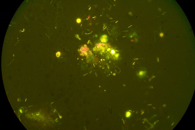
Photo: U.S. Geological Survey

Photo: Umberto Salvagnin
Other bacteria do not have flagella necessary for locomotion. But this does not mean that they will be motionless in the field of view of the microscope. No, it will seem to you that the bacteria are moving, all at once, like ants in a torn anthill. However, this is not an independent, active movement of the microbe, but the so-called Brownian movement.
The Brownian motion of any small particles floating in a liquid (by no means only microbes) is a consequence of the random thermal motion of the molecules of this liquid. Molecules put pressure on the particle from all sides, and it, so to speak, “marks time”.
But if bacteria are mobile under a microscope, then you will see how quickly they cross the field of view, freeze in place, and then again rush further. It is especially interesting to observe spirochetes, similar to a revived spiral from an electric stove. They are so thin that it is difficult to see a living spirochete under a normal microscope.
They are much better seen under a dark field microscope. You will probably find them in dental plaque; just take a good look - it is best to look for spirochetes during their movement. They either swim, wriggling like snakes, or twitch in place and even fold in half.
Live bacteria are not as convenient to view under a microscope as dead and stained ones.
With what magnification is it desirable to purchase a microscope in order to see microorganisms in the ACC?
Details of the structure of these organisms were studied precisely on stained preparations. To stain the bacteria, you need to put them on the glass (as they say, make a smear), dry it, heat it on a burner flame (so that the cells can be better painted later) and drop a drop of special paint on the smear.
If you find yourself in a microbiological laboratory, then, of course, there is a set of various paints. One of the most common is methylene blue. Since it is part of the ink for a fountain pen, for lack of a better one, you can sprinkle a drop of ink on a smear. After 6-8 minutes, the paint should be washed off with water and the smear dried.
Depending on which type of bacteria was stained, you will see balls or sticks under the microscope - straight, curved or comma-like. Chains can be formed from sticks and balls. The balls are sometimes grouped into groups of four, eight, and sixteen. Some sticks have thickenings at the ends, like a match head. These are the main forms of bacteria.
However, such a brief description is reminiscent of the words of a philosopher who defined man as a biped without feathers. In bacteria, even stained in the simplest way, one can find quite a lot of structural features. We will discuss some of these features here.
Rod-shaped bacteria are the most abundant in nature. The very word "bacteria" in Greek means "rod". One of the most common microbes, the so-called E. coli, has the shape of a long oval. E. coli lives in the large intestine; one gram of human feces can contain 2-3 billion of these microorganisms (imagine how many of them enter the external environment in a populated area!).
Pathogenic microbes, the causative agents of dysentery, typhoid, and paratyphoid, are indistinguishable from Escherichia coli in form. The causative agent of anthrax is also a stick, but with chopped off ends. Anthrax bacteria are often arranged in long filaments called chains.
The causative agents of tetanus, gas gangrene and many other diseases have the form of sticks.
Sometimes you can find the name "cholera comma". Indeed, the so-called vibrios are like a comma. These include the causative agent of cholera. Just don’t imagine the cholera comma in the form of a tadpole, as Mayakovsky liked to draw it in “Windows of GROWTH”. It is rather a curved stick of uniform thickness. Strictly speaking, this is not even a stick, but a segment of a spiral, one of its incomplete turns.
Globular bacteria are called cocci. Cocci, collected in clusters resembling grapes, are called staphylococci. Some of them, getting into wounds or scratches, cause suppuration and cause serious illness in young children.
A lot of misfortune is caused to a person by streptococci - microbes that look like strings of beads or a rosary. They cause erysipelas, tonsillitis, and even heart disease - endocarditis. Cocci, arranged in two - diplococci - a person owes diseases such as meningitis, pneumonia, gonorrhea.
It is easy to determine the shape of bacteria in a stained smear, but it is impossible to study the structure of a bacterial cell in all details. And if we still know a lot about the structure of bacteria, then this was helped by special methods of staining them and studying them under an electron microscope.
- microscopic method: light, phase-contrast, fluorescent, electronic;
- cultural method (bacteriological, virological);
- biological method (infection of laboratory animals);
- molecular genetic method (PCR - polymerase chain reaction)
- serological method - detection of antigens of microorganisms or antibodies to them;
Methods for preparing preparations for microscopy. With the help of a light microscope, microorganisms can be studied, both in a living and in a colored state. In the study of microbes in a living state, one can get an idea of the size, shape and nature of their movement. Sometimes shiny, strongly light-refracting granules and spores are visible inside a living cell. To study microbes in a living state, preparations of hanging and crushed drops are prepared. To prepare a hanging drop preparation (Fig. 19), a small drop of the test material suspended in a liquid (isotonic sodium chloride solution, meat-peptone broth) is applied to the center of the coverslip with a bacteriological loop. Then they take a special glass with a hole in the center and smear its edges with vaseline oil. Cover a drop of the test material on a cover slip with a slide hole so that the drop is in the center of the hole. Lightly press the glass slide and quickly turn it over. With proper preparation of the drug, the drop hangs down into the hole. Vaseline oil prevents it from drying out.
A crushed drop preparation is prepared by applying a drop of material suspended in liquid to a glass slide, which is then covered with a coverslip.
LIGHT OPTICAL MICROSCOPY
A microscope is used for light microscopy. an optical device that allows you to observe small objects. Image magnification is achieved by a system of lenses of the condenser, objective and eyepiece. A condenser located between the light source and the object under study collects light rays in the field of the microscope. The lens creates an image of the field of the microscope inside the tube. The eyepiece magnifies this image and makes it possible for the eye to perceive it.
Microscopy at home
The resolution limit of a microscope (the minimum distance at which two objects can be seen) is determined by the wavelength of the light and the aperture of the lenses. The theoretically possible resolution limit of a light microscope is 0.2 µm; real resolution can be increased by increasing the aperture of the optical system, for example, by increasing the refractive index. The refractive index (immersion) of liquid media is greater than the refractive index of air (“=1.0), several immersion media are used for microscopy: oil, glycerin, water. The mechanical part of the microscope includes a tripod, an object stage, macro- and micrometric screws, a tube, a tube holder.
Dark field microscopy allows the observation of live bacteria. For this, a dark-field condenser is used, which highlights the contrasting structures of the uncolored material. Before starting work, the light is installed and centered on the bright field, then the bright field condenser is removed and replaced with an appropriate system (for example, OI-10 or OI-21). The preparation is prepared according to the “crushed drop” method, making it as thin as possible (the thickness of the coverslip should not be thicker than 1 mm). The observed object appears as illuminated in a dark field. In this case, the rays from the illuminator fall on the object from the side, and only scattered rays enter the microscope lenses. Vaseline oil is suitable as an immersion liquid.
Phase contrast microscopy allows you to study living and unpainted objects by increasing their contrast. When light passes through colored objects, the amplitude of the light wave changes, and when passing through uncolored objects, the phases of the light wave change, which is used to obtain a high-contrast image in phase-contrast and interference microscopy. To increase the contrast, the phase rings are coated with a metal that absorbs direct light without affecting the phase shift. In the optical system of the microscope, a special condenser with a diaphragm revolver and a centering device is used; lenses are replaced with immersion apochromat lenses.
Polarizing microscopy allows imaging of unstained anisotropic structures (eg collagen fibers, myofibrils, or microorganism cells). The principle of the method is based on the study of an object in light formed by two beams polarized in mutually perpendicular planes.
Interference microscopy combines the principles of phase contrast and polarization microscopy. The method is used to obtain a contrasting three-dimensional image of unpainted objects. The principle of the method is based on the bifurcation of the light flux in the microscope; one beam passes through the object, the other - past it. Both beams are connected in the eyepiece and interfere with each other.
Luminescence microscopy. The method is based on the ability of some substances to glow when exposed to short-wave radiation. In this case, the emitted light waves are longer than the wavelength that causes the glow. In other words, fluorescent objects absorb light of one wavelength and emit light in another region of the spectrum. For example, if the inducing radiation is blue, then the resulting glow may be red or yellow. These substances (fluorescein isocyanate, acridine orange, rhodamine, etc.) are used as fluorescent dyes to observe fluorescent (luminescent) objects. In a fluorescent microscope, light from a source (an ultra-high pressure mercury lamp) passes through two filters. The first (blue) filter traps light in front of the sample and allows light of the wavelength that excites the sample to fluoresce. The second (yellow) delays blue light, but passes yellow, red, green light emitted by a fluorescent object and perceived by the eye. Usually, the studied microorganisms are stained directly or with the help of AT or lectins labeled with fluorochromes. Drugs interact with Ag or other ligand-binding structures of the object. Fluorescence microscopy has found wide application for visualization of the results of immunochemical reactions based on the specific interaction of AT labeled with fluorescent dyes with Ag of the object under study.


- The latest methods of teaching traffic rules
- How to draw pictures by numbers
- Do-it-yourself home digital microscope
- How to choose the right paint for drawing
- When is the best time to sunbathe?
- What kind of bird is better to have in an apartment?
- We put an apostille on the birth certificate on our own
- Is it possible to give flowers in pots - signs
- How to make cat ears
- Gray bag: what to wear and combine?
- How to get started with Faberlic: tips for new consultants
- Bioinsecticide Lepidocid: purpose, properties and application procedure Lepidocide waiting period
- How to change the language to Russian in steam
- Dendrobium noble: room care
- Morphology of plants general concepts - document
- Planting, propagation and care of bamboo at home, photo Growing bamboo from seeds
- How to strengthen the cellular signal for the Internet in the country
- Sanskrit reveals the forgotten meaning of Russian words (2 photos)
- The oldest language Sanskrit programming language of the future Dead language Sanskrit
- Who has dominion over all the earth?